Jayesh B. Shah, MD, CWSP, UHM, is the President of South Texas Wound Associates, PA, San Antonio, Texas, where he provides clinical wound care services in San Antonio and the surrounding communities. He is also the President of TIMEO2 Healing Concepts, LLC in San Antonio, Texas, which provides consulting and education services in wound care and hyperbaric medicine both nationally and internationally. His degrees include MBBS from M. S. University, India and MD in Internal Medicine from St. Luke’s Roosevelt Hospital, Columbia University, New York. He is Board Certified in Internal Medicine, Board Certified in Undersea and Hyperbaric Medicine, Certified in Wound Management, Certified in Hyperbaric Medicine, Past Chair of American College of Clinical Wound Specialists and Past Vice President of American College of Hyperbaric Medicine. He has been the recipient of the Enterpreuner of the year award by the Alamo Asia Chamber of Commerce (2016); the Paul James Sheffield Education Award for Lifetime Dedication to Education in the field of Undersea and Hyperbaric Medicine (2014); Jefferson C. Davis Memorial Award for Excellence in Clinical Hyperbaric Medicine (2007 and 2011); Carolyn Sue Award (2009); Young Scientist/Medical Doctor Award (2008); Community Service and Leadership award by Alamo Asian American Chamber of Commerce (2008). He has published 3 books- “Wound Care Certification Study Guide” in 2011 with its second edition 2016, Textbook of Clinical Wound Medicine: A Evidence Based Approach in press for release in 2017. He has authored over 40 chapters on various wound topics in 4 books in addition to 30+ scientific articles in wound care and hyperbaric medicine. As an Assistant Editor of the Journal of ACCWS, heregularly writes a column on certification exam in wound care.
Shah_Current Dialogues in Wound Management_2019_Volume 5_Issue 2
INTRODUCTION
Chronic wounds usually cost between $28 billion to $31.7 billion and affect 15% of 8.2 million Medicare beneficiaries.1 As chronic wounds are a prevalent and complex problem for wound care clinicians, an organized and systemic approach to their management is required.2 The TIMEO2 (tissue debridement, infection management, moisture control, edge effect, oxygen therapy) wound bed preparation principle has been elucidated by the author and represents a systemic approach to correct molecular and cellular abnormalities and is a critical component to promote the healing of chronic wounds.
Advanced wound therapies and dressings have evolved to help maintain appropriate levels of wound bed moisture, promote development of granulation tissue, and remove exudate and infectious materials. However, some wounds require the additional step of a skin graft. Traditional split-thickness skin grafts (STSGs) require an operating room, anesthesia, and cause the creation of a secondary wound at the donor site. However, epidermal grafts may provide an alternate option to STSGs.
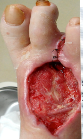
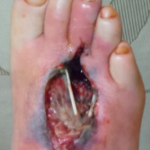 Figure 1. Wound at presentation
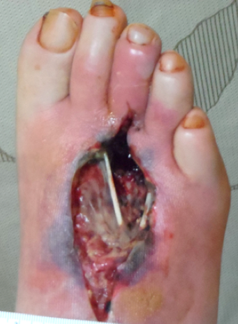
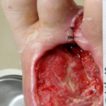 Figure 2. Wound after 2 weeks of treatment with 0.125% Dokin’s solution sooked gauze dressings
Epidermal grafts utilize autologous skin on the wound after the wound has already been prepared for expedited epithelization. In our experience, most chronic wound care patients prefer epidermal grafting in the clinic as there is no requirement for anesthesia and minimal to no complications at the donor site as only the epidermal layer is harvested. Also, in certain conditions like pyoderma gangrenosum and for anatomical locations like the plantar area, epidermal harvesting may be preferred over a traditional STSG.
This case study reports the use of advanced wound therapies and dressings for wound bed preparation followed by epidermal grafting to promote wound healing in a dorsal right foot ulcer
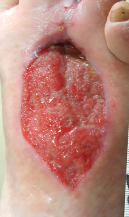
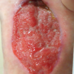 Figure 3. Wound after 1 month of VAC® Therapy and HBOT
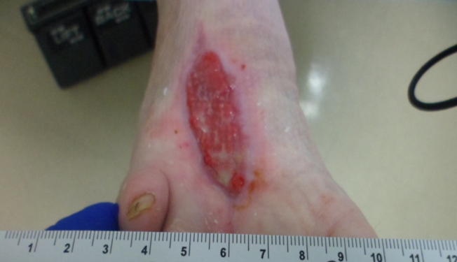
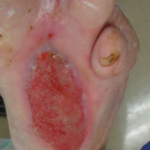 Figure 4. Wound 7 weeks after presentation.
CASE STUDY
This case study delineates that the patient requires all components of TIMEO2 for wound bed preparation
(T) Tissue debridement – where the patient requires multiple surgical debridement procedures
(I) Infection management – where the patient receives 6 weeks of intravenous ertapenem based on tissue and bone culture
(M) Moisture control with negative pressure wound therapy (NPWT)
(E) Edge effect with collagen
(O2) Correction of hypoxia with hyperbaric oxygen therapy (HBOT) for wound bed preparation. Once the wound bed is prepared with good granulation, the epidermal graft is used for expedited epithelialization
A 45-year-old male who works as an auto mechanic with a history of type II diabetes and hypertension presented with a wound on the dorsal right foot present for 50 days. The patient had been trying to manage the wound on his own with over-the-counter medications under the guidance of a primary care physician. However, the wound worsened, and the patient was admitted to the hospital with an infected deep abscess and severe diabetic foot infection. The wound underwent initial sharp debridement in the operating room. The patient was initially started on broad spectrum antibiotics (intravenous vancomycin and piperacillin/tazobactum)
The wound was classified as a Wagner Grade 3 ulcer stemming from a diabetic wound/ulcer of the lower extremity (Figure 1). The wound measured 10 cm length x 4 cm width x 3 cm depth with exposed muscle and tendon. There was a large amount of serosanguinous drainage noted with the wound margins undermined and no granulation within the wound bed. We treated the patient’s wound with 4×4 gauze soaked with 0.125% Dakin’s solution. A 6-week course of intravenous ertapenem was initiated. Wound cultures were positive for methicillin sensitive Staphylococcus aureus
After two weeks, the patient’s wound margin was flat and intact with granulation tissue (67-100%) observed in the wound bed (Figure 2). A small area (1-33%) of adherent slough was noted in the wound bed following another surgical debridement and third toe amputation. Hyperbaric Oxygen Therapy (HBOT) at 2 atmospheres absolute (ATA) for 90 minutes a day and V.A.C.® Therapy at -125 mmHg was initiated. Dressing changes occurred three times per week.
A month later, the wound measured 7.5 cm x 3.5 cm x 0.9 cm; there was ~0.5 cm of undermining from 9-11 o’clock (Figure 3). A large amount of serosanguinous drainage was observed. The wound margin was still flat and intact. V.A.C.® Therapy and HBOT was continued.
Seven weeks after presentation, the wound measured 7.3 cm x 3.0 cm x 0.5 cm (Figure 4). There was a minimal amount of serosanguinous drainage, presence of 67-100% granulation tissue, and a small area of loose slough (1-33%) observed within the wound bed. V.A.C.® Therapy and HBOT were discontinued and the wound was deemed suitable for epidermal graft placement.
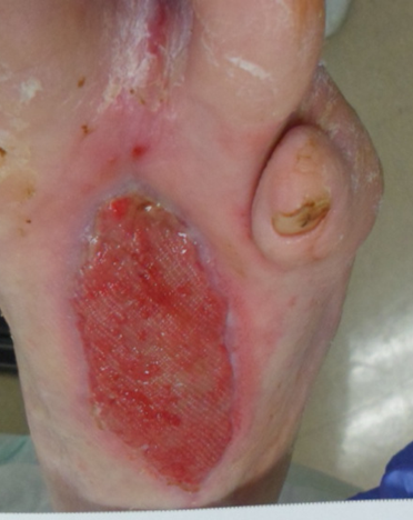
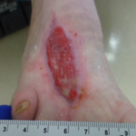 Figure 5. Wound one month after epidermal grafting
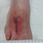 Figure 6. Wound completely epithelialized 18 weeks after presentation
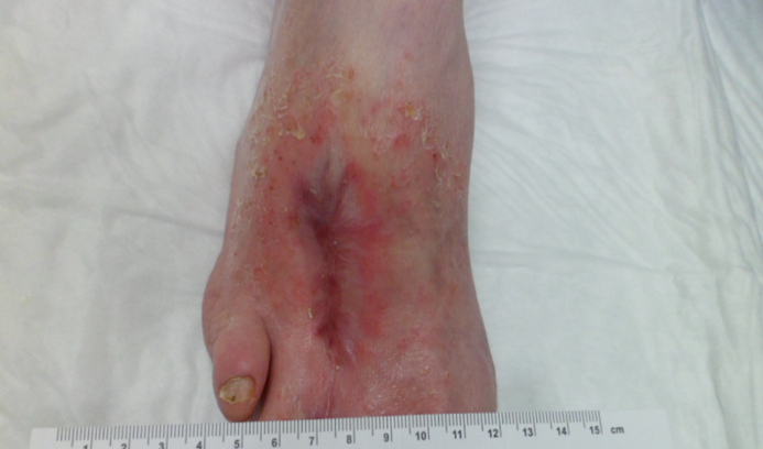
The patient’s right thigh was prepared for epidermal graft harvesting through hair removal and 70% isopropyl alcohol wash. The CELLUTOME™ Epidermal Harvesting System was applied to the thigh and epidermal grafts were harvested. The epidermal grafts were transferred to the wound using ADAPTIC TOUCH™ Non-Adhering Silicone Dressing. The epidermal grafts were bolstered with a secondary dressing, which was changed once a week, according to manufacturer’s instructions. A transparent film dressing was applied to the donor site. The donor site was fully healed 7 days after graft harvesting. One month after epidermal grafting, the wound continued to improve and measured 5.4 cm x 1.8 cm x 0.3 cm. The ADAPTIC TOUCH™ Non-Adhering Silicone Dressing and secondary dressings were discontinued. A hydrogel and collagen dressing were applied with dressing changes once per week, according to manufacturer’s instructions.Approxmately 18 weeks after presentation the wound was completely epithelialized (Figure 6).
CONCLUSION
Management of chronic wounds is a dynamic process, which requires evaluation and assessment of the wound on a regular basis. A comprehensive wound management algorithm using the TIME02 principle may help to prepare the wound bed. It is also important to understand patient-specific factors and social determinants of health which may also affect wound healing. Ulcers with a poor prognosis should be treated with a more advanced therapy. It is important to identify patients who are unlikely to respond to conventional care early so that advanced therapies can be considered and initiated. Application of epidermal grafts is a good alternative treatment over traditional split-thickness skin grafts once the wound has 100% granulation tissue. Epidermal grafting has shown promising results after wound bed preparation using advanced dressings in a variety of wounds including those with numerous patient comorbidities and in patients where a STSG is contraindicated.
References
1.Nussbaum SR, Carter MJ, Fife CE et al. An Economic Evaluation of the Im-pact, Cost, and Medicare Policy Implications of Chronic Nonhealing Wounds. Value in Health 2018;21(1):27-32. doi:10.1016/j.jval.2017.07.007.
2.Fife C, Shah JB. Wound Bed Preparation and Advanced Technologies for wound healing. In: Shah JB, Sheffield PJ, Fife CE, eds. Wound Care Certi-fication Study Guide. 2nd ed. North Palm Beach, FL: Best Publishing Com-pany; 2016:97-112.
3.Woo K, Beeckman D. Wound Bed Preparation. In: Shah JB, Sheffield PJ, Fife CE, eds. Textbook of Chronic Wound Care. 1st ed. North Palm Beach, FL: Best Publishing Company; 2018:127-147.
4.Shah JB. Correction of Hypoxia, a Critical Element for Wound Bed Prepa-ration Guidelines: TIMEO2 Principle of Wound Bed Preparation. Journal of the American College of Certified Wound Specialists 2011;3(2):26-32. doi:10.1016/j.jcws.2011.09.001.
5.Serena TE. Use of epidermal grafts in wounds: a review of an automated epidermal harvesting system. J Wound Care 2015;24(4 Suppl):30-34. doi:10.12968/jowc.2015.24.Sup4b.30.
6.Ramos JV, Schmidt M, Wu SC. Epidermal skin grafts for the treatment of chronic lower extremity ulcers. Podiatry Management 2013;95-96, 98-100, 102, 104.
7.Serena T, Francius A, Taylor C, Macdonald J. Use of a novel epidermal harvesting system in resource-poor countries. Adv Skin Wound Care 2015;28(3):107-112.
8.Richmond NA, Lamel SA, Braun LR, Vivas AC, Serena T, Kirsner RS. Epi-dermal grafting using a novel suction blister-harvesting system for the treatment of pyoderma gangrenosum. JAMA Dermatology 2014;150(9):999-1000.
Photos and patient information courtesy of Jayesh Shah, MD, South Texas Wound Associates, PA, San Antonio, TX.As with any case study, the results and outcomes should not be interpreted as a guarantee or warranty of similar results. Individual results may vary depending on the patient’s circumstances and condition.NOTE: Specific indications, contraindications, warnings, precautions, and safety information may exist for Systagenix and KCI (Acelity companies) products. Please consult a healthcare provider and production instructions for us prior to application.
