
D. Heath Stacey, MD is a plastic surgeon in private practice in Northwest Arkansas. His clinical interests include breast reconstruction, complex abdominal reconstruction, lower extremity reconstruction, breast and body contouring, and facial aesthetic surgery.
Stacey_Current Dialogues in Wound Management_2016_Volume 2_Issue 3
NOTE: As with any case study, the results and outcomes should not be interpreted as a guarantee or warranty of similar results. Individual results may vary depending on the patient’s circumstances and condition.
Wounds to the lower extremity resulting from trauma cause significant morbidity often requiring multiple operations and prolonged time to healing. In this article, we will review the importance of serial debridement, management of these difficult wounds, and a representative case will be presented.
These wounds are complex and often have components of crush, avulsion and lacerations to the soft tissue. Additionally, there are often concomitant fractures present which will need to be addressed before definitive soft tissue coverage.
Medina et al provide a good overview of evidence-based management of lower extremity trauma wounds. The decision whether to perform an early amputation or attempt limb salvage is one often encountered early in severe injuries. Injury scoring systems may provide some usefulness, but do not help with all patients. In patients with severe vascular or nerve injuries in conjunction with large areas of soft tissue loss > 10 cm and large areas of exposed, comminuted fractures, early amputation should be considered. Also, patient co-morbidities such as diabetes mellitus, peripheral vascular disease, and other injuries resulting from the trauma should be considered when deciding on pursuing limb salvage.
If limb salvage and reconstruction are to be the treatment course, several treatment goals are identified. Many of these wounds present with heavy contamination, a large crush component, and /or multi-level injury. Fixation of any fractures with external fixators and serial debridements are the first goals of treatment. Surgical debridements are performed as many times as necessary, usually 2-3 days apart to remove devitalized and contaminated soft tissue. It is also necessary to remove any bone that is potentially compromised or devascularized as this later can be a source of late osteomyelitis.
Use of negative pressure wound therapy on the wound(s) between debridements should be considered in order to stimulate granulation tissue and help control bacterial contamination. V.A.C. VERAFLO™ Therapy is often used as well in contaminated wounds which include industrial accidents, farm injuries, or wounds with a large crush / avulsion component. Parrett et al showed in their series of patients that this negative pressure wound therapy may help with bridging before definitive closure is obtained.
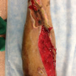 Figure 1a. Left leg upon presentation
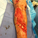 Figure 1b. Left leg post debridement.
CLINICAL CASE SCENARIO
The patient in Figure 1 presented as a 46 y/o gentleman with no significant past medical history who developed a significant multi-level lower extremity injury after a tree he was cutting down fell on his left lower extremity. He was referred by the orthopedic surgeon for soft tissue closure. His external fixator had been removed and an intramedullary rod had been placed in his tibia. Figure 1 shows his leg with some granulation tissue present. The eschar distally was concerning as this area was overlying tibia on the middle third of the leg. After debridement, the bone was exposed.
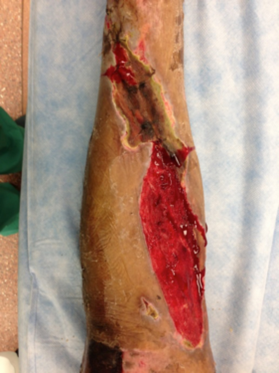
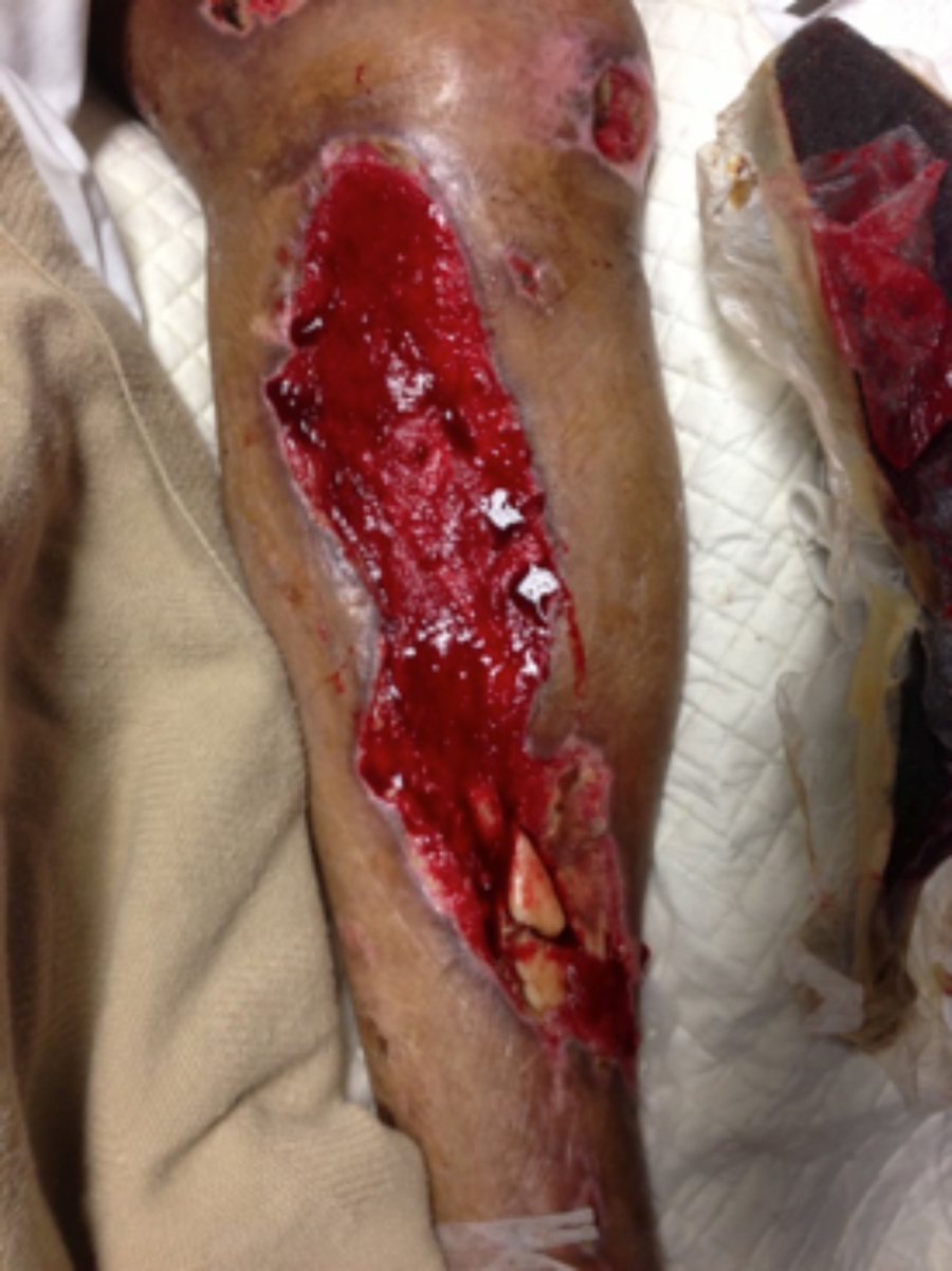 Figure 2. Left leg during NPWT
Figure 2 shows the patient’s leg after NPWT for 1 week. There are signs of healing, but exposed fragments of tibia remain distally. A debridement was performed at this point and
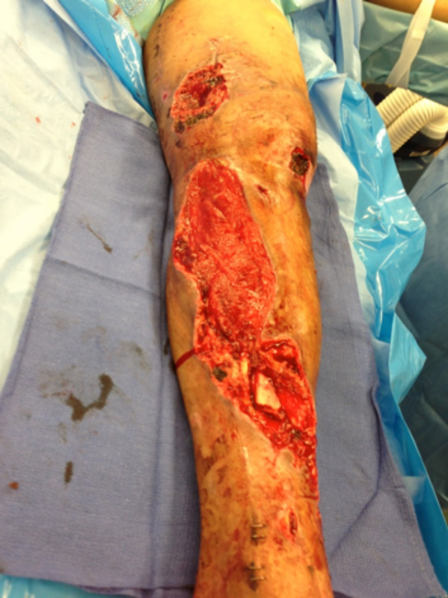
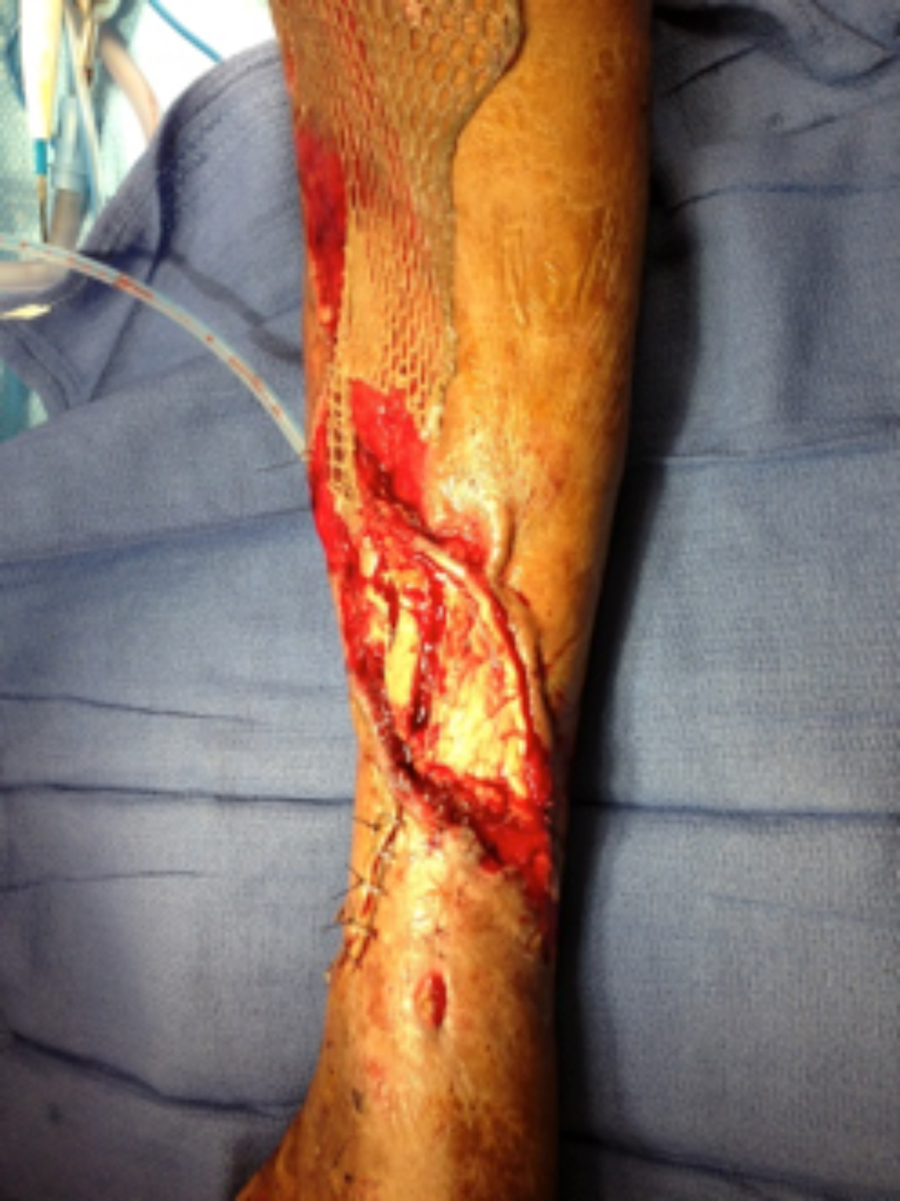 Figure 3. Left leg at time of muscle flap and skin graft
NPWT was continued for 1 more week.
Figure 3 illustrates the patient’s defect, which has now been covered with a hemi-soleus muscle flap to cover the tibia and split thickness skin grafting to cover the granulated muscle bellies. This patient achieved wound closure (Figure 4) and is ambulating.
The importance of removing all devitalized tissue before definitive closure is illustrated in this case. Also, the negative pressure therapy is a critical component of management between debridements and before final soft tissue coverage.
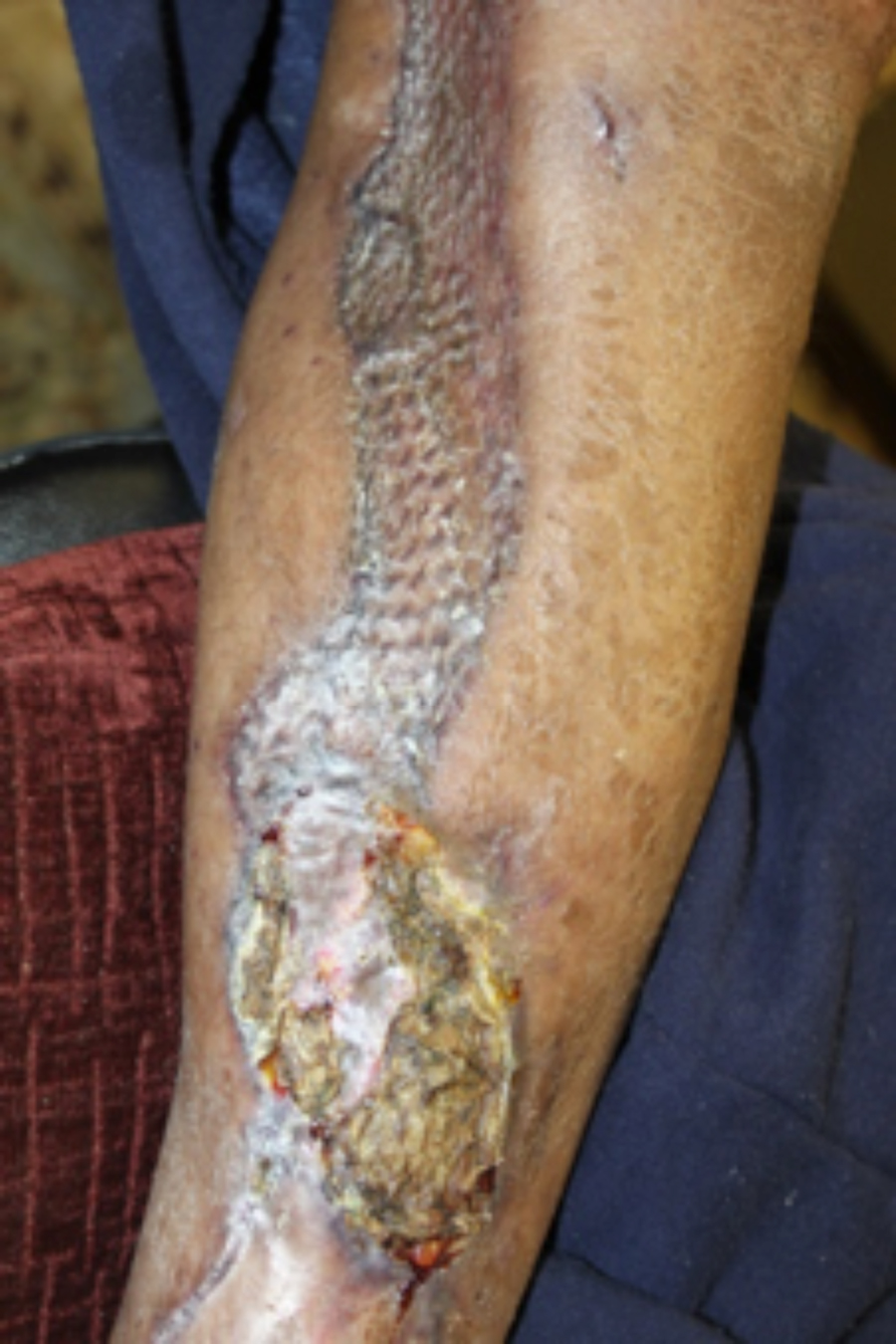 Figure 4. Left leg after wound flap and graft healing
References
1.Medina ND, Kovach SJ III, Levin LS. An evidence-based approach to lower extremity acute trauma. Plastic and Reconstructive Surgery 2011;127.2:926-931.
2.Parrett BM, et al. Lower extremity trauma: trends in the management of soft-tissue reconstruction of open tibia-fibula fractures. Plastic and Reconstructive Surgery 2006;117.4:1315-1322. 2013;132:1569.
3.Fluieraru S, et al. Sterile-water negative pressure instillation therapy for complex wounds and NPWT failures. J Wound Care. June 2013;22(6).
4.Gabriel A, et al. Negative pressure wound therapy with instillation: a pilot study describing a new method for treating infected wounds. Int Wound J. 2008;5:399–413.
