
Kersten Reider received her Diploma in nursing from the Reading Hospital School of Nursing and then went on to finish her BSN at Alvernia University. She is currently enrolled in the Adult-Geriatric Nurse Practitioner Program at Bloomsburg University. She is a Certified Wound, Ostomy, Continence nurse who has been working in wound care for the past six years in a large Acute Care Hospital in Reading Pennsylvania.
Reider_Current Dialogues in Wound Management_2016_Volume 2_Issue 3
NOTE: As with any case study, the results and outcomes should not be interpreted as a guarantee or warranty of similar results. Individual results may vary depending on the patient’s circumstances and condition.
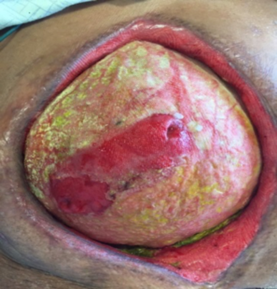 Figure 1. Abdominal Wound (27x17x8cm) at presentation with two visible enteroatmospheric fistulas
ABSTRACT
Background: Fifty-four year old morbidly obese female admitted with a small bowel obstruction and a large ventral hernia. The patient, who underwent an exploratory laparotomy with lysis of adhesions and ventral hernia repair with mesh placement, ultimately developed an enteroatmospheric fistula (EAF). Several months of hospitalization were required to manage the wound and contain effluent from the fistula effectively.
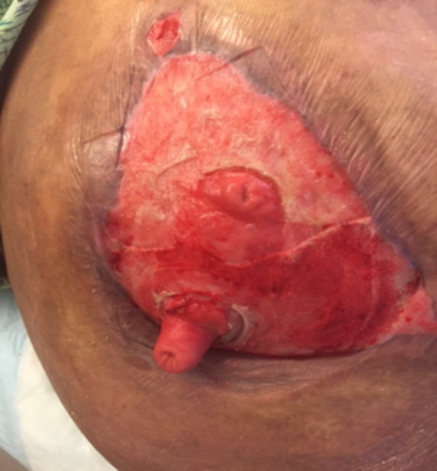
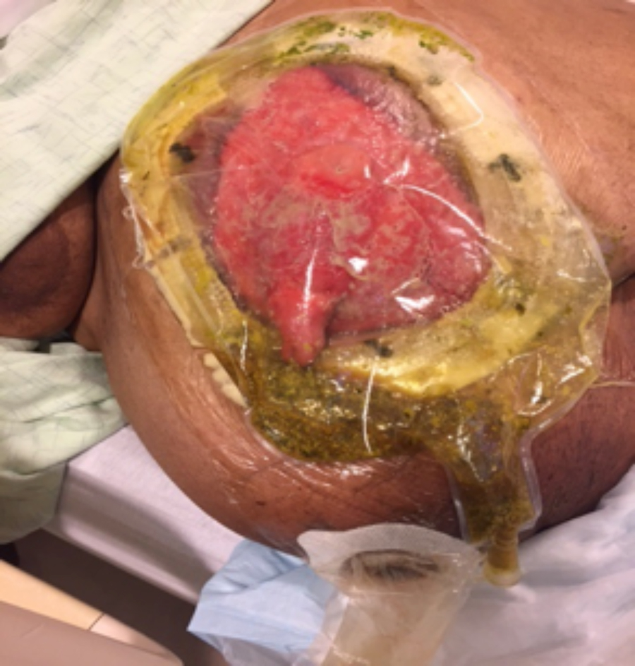 Figure 2. Wound with fistula management pouch to contain effluent
Methods: A fistula management pouch was utilized for several months to encompass the wound and contain effluent. This method proved to be ineffective. The fistula was then isolated utilizing a collapsible enteroatmospheric fistula isolation device and an ostomy appliance to contain effluent. The wound was then managed with negative pressure wound therapy (NPWT).
Conclusion: The application of the collapsible enteroatmospheric fistula isolation and effluent containment devices in conjunction with NPWT produced positive outcomes for this patient, including patient satisfaction, decreased financial burden, promotion of wound healing, and ultimately wound closure.
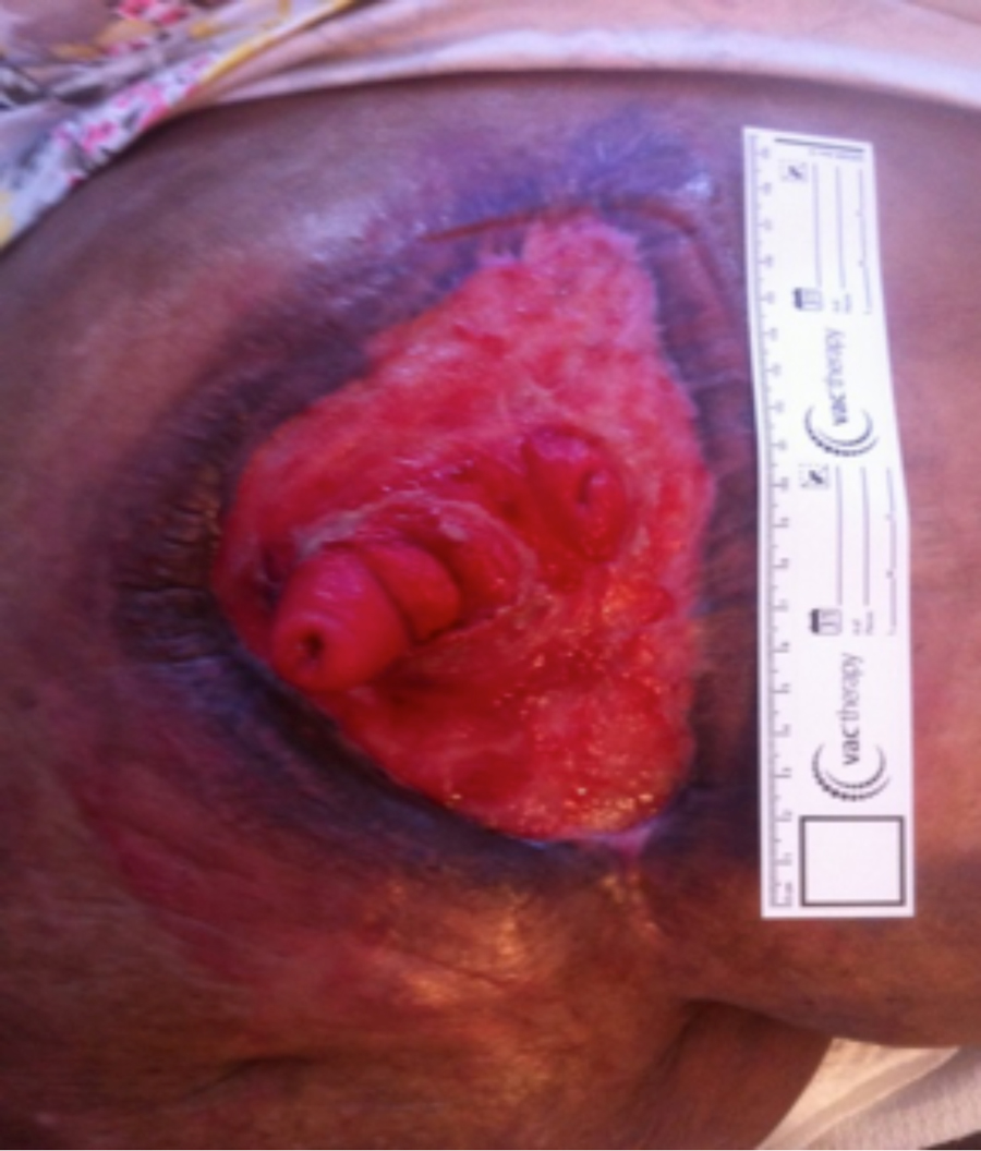
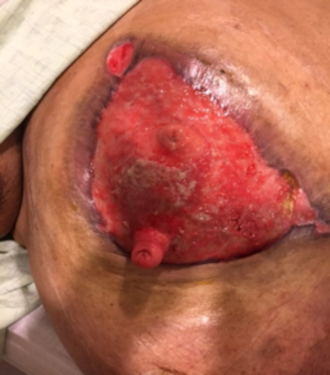 Figure 3. Wound (25x16x2cm) after pouch removal
INTRODUCTION
Enteroatmospheric fistulas (EAFs) are a common complication of open abdomen bowel surgery with an estimated 2%-50% of patients developing fistulas post- surgery.1The presence of an EAF increases patient morbidity and provides a challenge to the wound care team.2 Traditionally, management of EAFs has involved placing large fistula management pouches over the wound to collect and contain effluent. However, more recent techniques isolate the fistula and apply negative pressure wound therapy (NPWT) to the rest of the wound.2 Isolation of the EAF and application of NPWT to a large abdominal wound proved effective in the promotion of granulation tissue formation and decreasing wound dimensions.
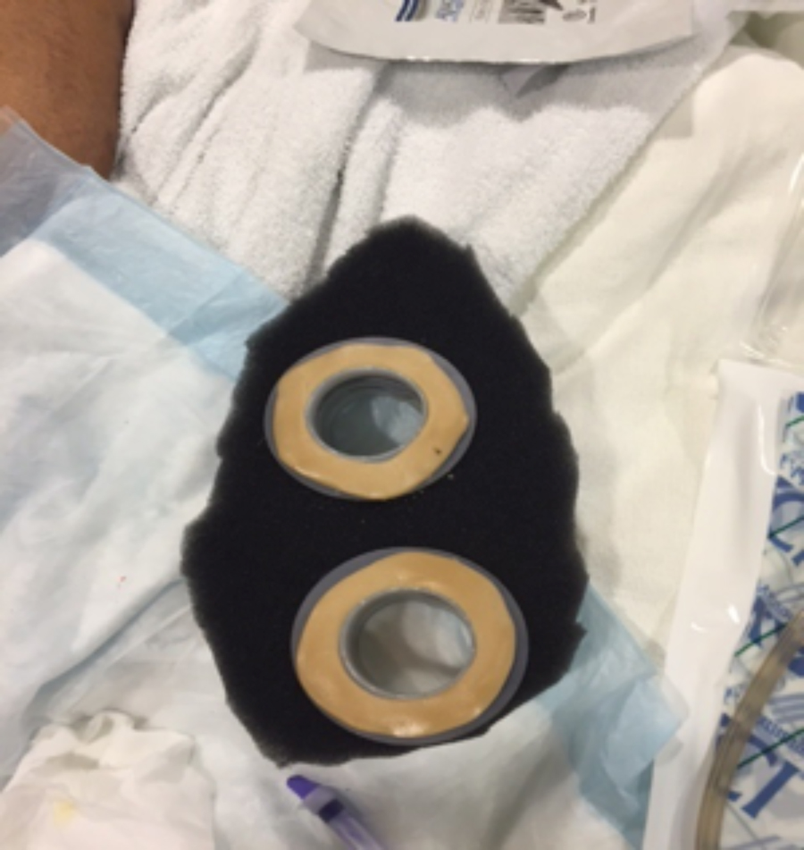 Figure 4. Negative pressure polyurethane foam with collapsible enteratmospheric fistula isolation devices and barrier rings
CASE
A fifty-four year old morbidly obese female, who had undergone abdominal wall hernia repair in 2007, presented to the Emergency Department with abdominal pain, nausea and vomiting, and no bowel movement for four days. An obstruction series revealed a partial versus full complete bowel obstruction. At this time, the decision was made to proceed with a CT scan of the abdomen, place a nasogastric tube (NGT) for decompression, start IV fluid hydration and admit the patient to a medical-surgical unit for further evaluation from the surgical team. The CT results indicated there was a large ventral wall hernia complex containing multiple small bowel loops. The hernia measured 35 x 12.5 x 29cm. The distal colon was collapsed, and the findings were consistent with a small bowel obstruction. Consultation by the surgical team recommended conservative non-operative management. Over the course of nine days, the patient was started on total parental nutrition, the NGT was left in place for decompression, and eventually a clamping trial was initiated, and a bariatric surgery consultation was completed.
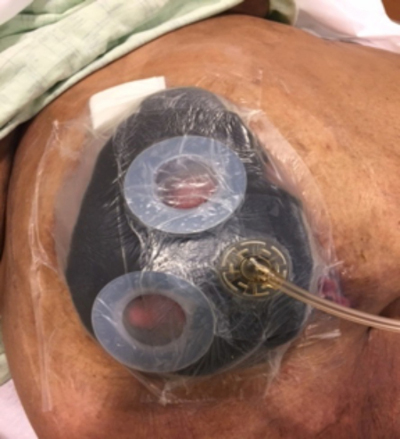 Figure 5. Application of NPWT with polyurethane foam and the use of the collapsible enteroatmospheric fistula devices to isolate the fistulas
Unfortunately, the patient failed conservative treatment and surgical intervention became inevitable. The patient underwent an exploratory laparotomy, extensive lysis of adhesions, explantation of previously placed mesh, open appendectomy, incisional hernia repair with mesh, and bilateral salpingo-oophorectomy. During the operative procedure a piece of Vicryl® (Ethicon, Inc.) mesh was used to cover all of the bowel that was reduced into the true abdomen. A piece of Bio-A® (W.L. Gore & Associates) mesh was sutured along the rectus muscles to provide a second buttressing for the hernia repair. Primary closure was completed and the patient was transferred to the surgical intensive care unit. For several weeks following surgery the patient struggled with persistent fevers, tachycardia, decreasing urine output, increasing creatinine and ventilator dependent respiratory failure.
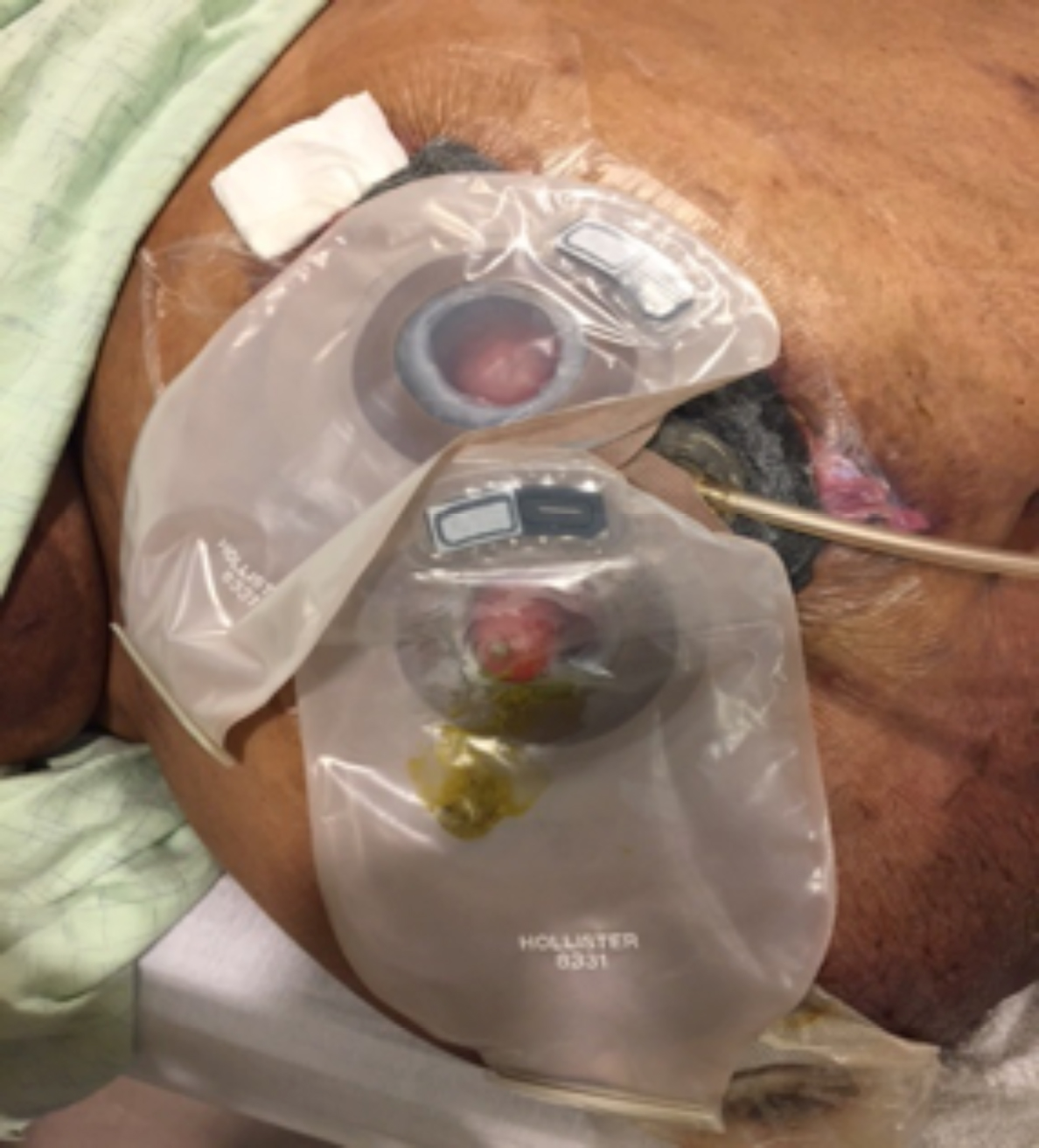 Figure 6. Ostomy appliances placed over the collapsible enteroatmospheric fistula isolation devices to collect effluent while NPWT in place
Two weeks post-operatively a JP drain that was placed in the abdomen was noted to contain thick drainage with increased volume that made the surgical team question the development of a fistula. The following day the patient underwent abdominal wound exploration and washout with findings consistent with a small bowel fistula with active drainage. In an attempt to contain drainage, the fistula was intubated with a red rubber catheter and NPWT was applied to the wound. At the request of the surgeon, a consult was placed to the Wound, Ostomy, Continence Nurse (WOCN) to manage the NPWT and to provide regular wound dressing changes.
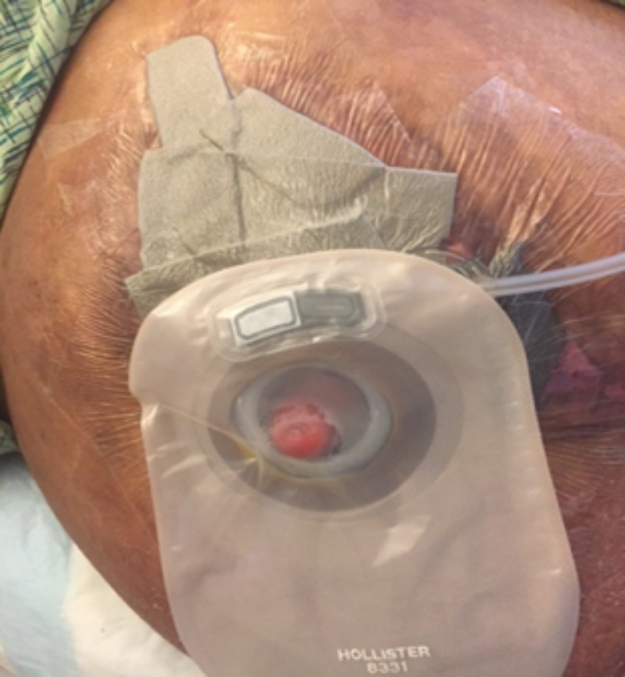 Figure 7. Silver foam dressing to superior aspect of the wound, black polyurethane foam to the inferior aspect of the wound with fistula isolation device and ostomy appliance
Four days post-operatively the abdominal dressing was changed at the bedside in collaboration with the surgeon. During the dressing removal, the red rubber catheter that intubated the fistula became dislodged and a large amount of brown drainage was noted
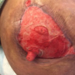 Figure 8. Abdominal wound four months from presentation (21x13x0.5cm) at discharge to home
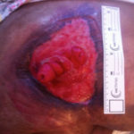 Figure 9. Abdominal wound six months after presentation (14×11.5×0.4cm)
pooling in the inferior aspect of the abdominal wound along with the right lateral gutter. At this time a collaborative decision was made to discontinue NPWT and apply a dressing that would contain the large amounts of effluent emanating from the fistula. The wound was cleansed with saline. Vaseline impregnated mesh was placed against wound base followed by a layer of saline moistened roll gauze. An 18 French red rubber catheter was placed along
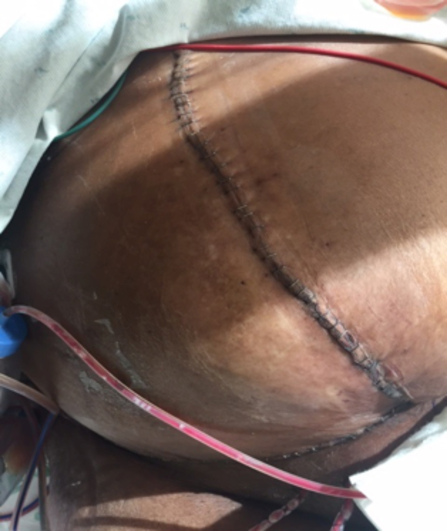
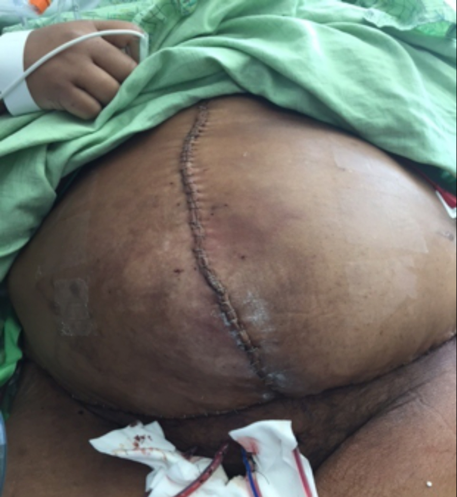 Figure 10. Abdominal wall reconstruction and closure seven months after presentation
the wound bed, the wound depth was filled with saline moistened roll gauze and then covered with an occlusive dressing. The red rubber catheter that was placed into wound bed was connected to low continuous wall suction. The dressing changes were performed three times a week. This particular treatment plan ideally kept the drainage contained, which prevented skin breakdown from the caustic fistulous output and was less labor intensive for the bedside nurses than dressing changes every eight hours. This method of treatment was continued for approximately a month.
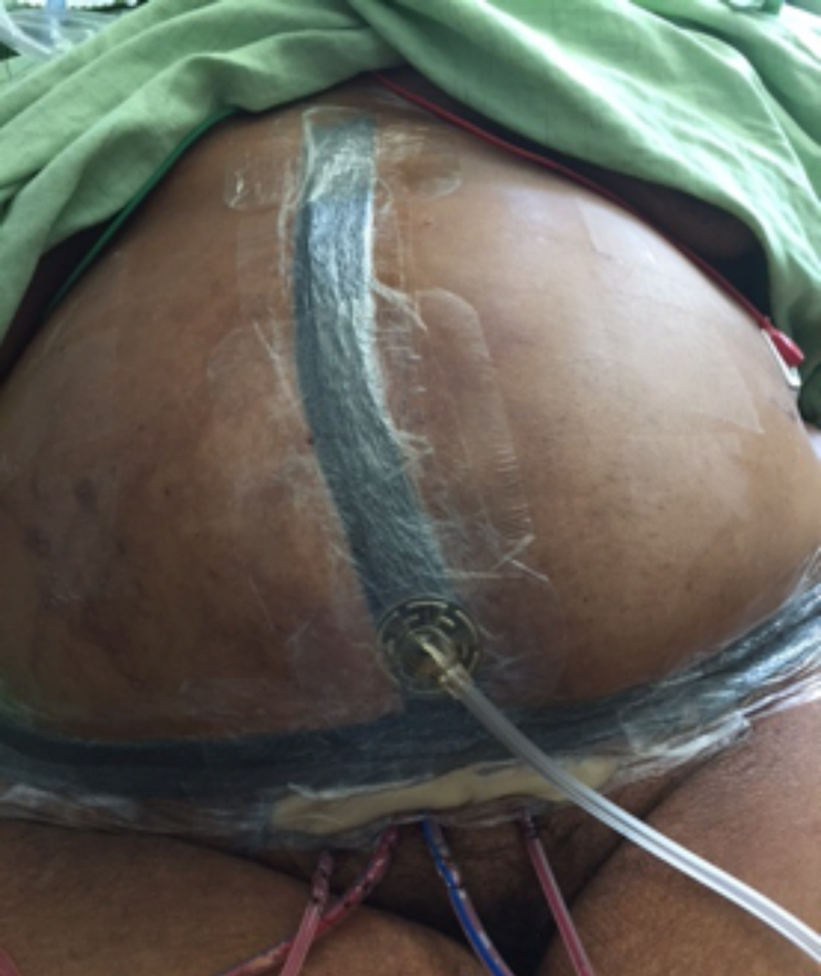 Figure 11. Incisional NPWT for five days postoperatively
As the patient progressed medically, she started to consume a general diet and began to ambulate short distances, which caused the treatment plan to become ineffective. The wall suction was unable to pull the thicker effluent, which in turn caused frequent leaking issues and multiple dressing changes. This treatment plan was also not a feasible option for discharge. Failure of the current treatment plan led the WOCN to consider other options that once again would contain drainage and ultimately be appropriate for discharge.
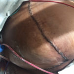 Figure 12. Incision after removal of incisional NPWT
 Figure 13. Silver Silicone Incisional Border dressing for discharge home
Two months had passed since surgery, and the patient was medically progressing towards discharge. Unfortunately, the current treatment plan for fistula management was not a feasible option. Assessment of the wound revealed that the edges had contracted slightly and the fistula effluent had thickened and decreased in volume. At this point, the decision was made to apply a large fistula management pouch that would encompass the wound and contain the fistula output. This method of treatment was continued for a few weeks, with the pouches being changed three times a week. Proving to be a successful treatment option, the patient was discharged to a skilled nursing facility. The patient was not in favor of residing at a skilled nursing facility, but due to the complexity of the wound care and the need for rehabilitative services, this was the most appropriate option.
During the patient’s three-month stay at the skilled nursing facility she encountered many complications. The patient was sent to the emergency room six times for abdominal pain along with nausea and vomiting. The WOCN had been consulted during these emergency room visits to evaluate the patient for a leaking fistula management pouch. Per the patient, the fistula management pouch had been leaking daily, sometimes causing the pouches to be changed multiple times a day. Even though her wound was contracting slowly, the periwound was beginning to breakdown. The patient expressed frustration in her current treatment plan along with feeling depressed. She wanted to be home with her family and participate in activities such as bingo and spend time with her grandchildren.
Eventually the patient’s abdominal pain along with nausea and vomiting was accompanied by a fever. She was admitted to an inpatient medical-surgical unit for further evaluation and workup. The fistula management pouch was removed to assess the wound. The wound measured 25x16x2cm. There were two visible fistulas in the wound base. The inferior fistula was protuberant and functioning for high output of effluent, while the superior fistula was flush and appeared nonfunctioning. The wound base had minimal granulation tissue. The periwound was denuded with ulcerations secondary to leakage of effluent. After assessment, it was determined that a better treatment plan needed to be implemented.
The treatment plan needed to focus on the promotion of wound healing along with the containment of effluent. The goal was to isolate the fistula and contain effluent while utilizing negative pressure to promote granulation tissue and wound contraction. A collapsible fistula isolation device (Wound Crown®, Fistula Solutions Corporation) was used to isolate the fistula while allowing effluent to be collected in an ostomy appliance while the wound was treated with NPWT.3 The flexible device is designed to create a channel for effluent while maintaining the integrity and benefits of the NPWT dressing.3 The base of the fistula isolation device was cut to contour the patients wound bed and fit over the fistula. A barrier ring was then applied to the base of the fistula isolation device to help achieve a seal between the wound bed and the device. The negative pressure polyurethane foam was then measured and cut to fit into the wound bed. After tailoring of the foam, a 4cm hole was cut into the foam where the fistula would be centered. The collapsible fistula isolation device was then inserted into the hole created in the polyurethane foam until the top and bottom flange lay flush against the foam. The flanges anchored the collapsible fistula isolation device within the polyurethane foam and created a channel to capture the effluent draining from the fistula. The assembled dressing was then
placed into the wound bed so the collapsible fistula isolation device base was centered over the fistula. Once centered, the device and foam were covered by clear drape and NPWT at -125mmHg continuous. After a seal was achieved, an opening was cut in the clear drape at the top of the fistula isolation device. An ostomy appliance was applied to the top flange of the fistula isolation device to contain effluent. The entire dressing was changed three times a week by the WOCN.
The new treatment plan with the collapsible fistula isolation and NPWT proved to be successful with some fine tuning. During the initial application of the collapsible fistula isolation device and NPWT, the fistula isolation device was applied to both superior and inferior fistulas. It was noted that the superior fistula was nonfunctioning; therefore, Vaseline impregnated gauze was applied over that fistula followed by NPWT. Only the inferior fistula continued to be isolated. After one week of NPWT being applied, the superior aspect of the wound had granulated flush with periwound skin, but there was still notable depth at the inferior aspect of the wound. The decision was made to apply a silver silicone foam dressing to the superior aspect of the wound to promote healing, but prevent hypergranulation tissue from forming. NPWT was continued to the inferior aspect of the wound along with isolating the fistula with the collapsible fistula isolation device. This treatment continued to be successful. During the last dressing change prior to the patients discharge her wound measured 21 x 13 x 0.5cm. The wound base had significant amount of granulation tissue, the wound dimensions had significantly decreased, and the effluent from the fistula was contained. Due to the success of the new treatment plan, the patient was able to return home with visiting nurses instead of residing at a skilled nursing facility.
Seven months after presentation of an abdominal wound with enteroatmospheric fistulas the patient underwent abdominal reconstructive surgery. The procedure performed included takedown of the enteroatmospheric fistula, abdominal wall reconstruction, and a panniculectomy. The abdomen was closed primarily with staples. Post-operatively NPWT was applied to the incision line and kept in place for five days. This method of NPWT was to assist with cooptation of the incision line along with decreasing the incidence of post-operative surgical site infection. On the fifth day the NPWT dressing was removed. The incision was intact with no drainage or clinical signs or symptoms of infection. After removal of the NPWT dressing, a silver silicone bordered dressing was applied to the incision line in anticipation of discharge within the next few days. Successfully, the patient was discharged home with a closed abdomen.
References
1.Becker HP, Willms A, Schwab R. Small bowel fistulas and the open abdomen. Scandinavian J Surg. 2007;96:263-271.
2.Timmons J, Russell F. The use of negative-pressure wound therapy to manage enteroatmospheric fistulae in two patients with large abdominal wounds. Intl Wound J. 2014;11:723-729.
3.Heineman J, Garcia L, Obst M, et al. Collapsible enteroatmospheric fistula isolation device: A novel, simple solution to a complex problem. J Am Coll Surg. 2015;221(2):e7-e14.
4.Majercik S, Kinikini M, White T. Enteroatmospheric fistula: From soup to nuts. Nutrition in Clinical Practice 2012;27(4):507-512.
5.Terzi C, Egeli T, Canda A, et al. Management of enteroatmospheric fistulae. Intl Wound J. 2014;11(suppl 1):17-21.
6.Schecter W, Hershberg A, Chang D, et al. Enteric fistulas: Principles of management. Am Coll Surg. 2009;209(4):484-491.
