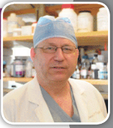
Dr. Orgill is Vice Chairman for Quality Improvement in the Department of Surgery at Brigham and Women’s Hospital and Professor of Surgery at Harvard Medical School. He is a reconstructive plastic surgeon and has a PhD from MIT in Medical Engineering. He is the Director of the Brigham and Women’s Hospital Wound Care Center and runs a tissue engineering and wound healing Laboratory. His lab at BWH is working to develop better technologies to treat wounds including work with artificial skin, micromechanical forces, platelets and stem cells. He has consulted for several medical device and start-up companies and is the inventor on several patents. He worked on the team that developed Integra®, a skin replacement therapy that has been commercially developed and used successfully on thousands of patients.
Orgill_Current Dialogues in Wound Management_2017_Volume 3_Issue 1
Lower extremity trauma continues to challenge surgeons to provide functional and aesthetic outcomes for our patients while minimizing multiple operations and chronic pain. Deciding between amputation and reconstruction can be a complex issue as both our ability to reconstruct limbs as well as prosthetic technologies have dramatically improved over the last 10 years. We will focus here on improvements in reconstructive products and techniques.
The most challenging soft tissue requirements occur in the distal tibial, ankle or foot where there is a relative lack of local soft tissues to transfer. There have been several advances in both products and techniques that now afford surgeons many more options when dealing with complex lower extremity trauma patients.
Marco Godina, a plastic surgeon from Ljubljana Yugoslavia, revolutionized lower extremity trauma treatment with his seminal paper published in September of 1986 following his untimely death earlier that year.1 He reviewed 532 patients with lower extremity microsurgical reconstruction and showed dramatically better results in patients treated within 72 hours of their injury that included a radical debridement and free tissue transfer. In this group, there was only one flap failure in 134 patients. Many aspects of both plastic surgical and orthopedic treatments have changed since that time, including the length of hospitalization which ranged from an average of 27 days in the immediate group to 156 days for those treated between 3 days and 3 months. We will review some of these changes in soft tissue management including both products and procedures.
PRODUCTS
NPWT
The advent of NPWT has changed how these wounds are managed and most commonly lower extremity traumatic injuries are initially treated with debridement followed by some sort of NPWT treatment. Newer devices are smaller, easier to use and can be used on either an intermittent or continuous modes. Other devices allow the irrigation of fluids within the interface materials. The advent of NPWT has in our institution been a major contributing factor to lowering the number of free flaps that we currently do for lower extremity trauma.2 These devices evacuate fluid, create an environment to promote wound healing, and keep the wound covered to prevent contamination from the external environment. For large wounds that need some sort of flap coverage, NPWT should not delay coverage. In our institution we try, when possible to close these wounds within a week of injury.
Scaffolds
Scaffolds allow host tissues to grow into a biological matrix. There are a number of scaffolds on the market or in development that are designed to replace a variety of structures including the dermis and peripheral nerves. They can be divided into semi-synthetic, such as porous collagen-GAG scaffolds or those derived from tissue that is subsequently decellularized. Typically, these scaffolds are either taken from human cadavers or processed from animal skins; e.g. porcine or bovine. Over time, scaffolds are infiltrated by cells and blood vessels from the wound. In areas where bone, joint or tendon is exposed, there is a possibility of bridging over these structures with vascularized tissue. The exact size of the defect that can be bridged depends on the condition of the wound, the age of the patient and co-morbid conditions. Being able to generate a dermis in the hostile environment of an acute wound can often allow the surgeon to perform a skin graft rather than doing a more extensive flap procedure.3
Placental Derived Materials
Placental derived materials have been used for many years to treat burns and other difficult wounds. The placenta is made of two distinct layers: the amnion and chorion. These layers are rich in growth factors that stimulate wounds to heal and contain abundant extracellular matrix (ECM) and well-preserved endogenous growth factors. New advances in processing techniques allow these materials to be made into dehydrated sheets with a long shelf life. Pure amniotic membranes have been quite useful in treating corneal injuries. For wound care, many products today use some combination of the amnion and chorion – both layers are quite rich in growth factors. These products have been shown to be effective particularly in diabetic foot infections.4
PROCEDURES
Improvements in Skin Grafting
Skin grafting has become more reliable as we learn how to better prepare the wound, treat the donor site and better affix the graft to the wound.5
Fibrin glue has shown to be quite effective in increasing skin graft take in difficult areas. Using a NPWT device as a bolster has also been studied in randomized controlled clinical trials to improve the take compared to a conventional bolster. Webster et al. conducted a comprehensive systematic review of the literature including trials involving people of any age, and in any care setting, involving the use of NPWT for surgical wounds. Included in this review were five eligible trials with a total of 280 participants. Two trials involved skin grafts and three involved acute wounds.6
Perforator Flaps
Our understanding of the anatomy of perforators based on basic work of Taylor7 has shown that flaps can be designed on single perforators that go through the skin. For small areas of exposed bone, these can be very helpful in covering the bone and skin grafts can be used to cover other areas of soft tissue defect.
Supermicrosurgery
Microsurgery has advanced so that blood vessel anastamoses can be reliably performed on blood vessels less than one mm in diameter. This allows the possibility of moving small areas of tissue to distal sites. This has been particularly useful in some diabetic patients with non-healing ulcerations on the foot.7
CONCLUSIONS
Better surgical products combined with better surgical techniques have given surgeons many more options for dealing with lower extremity traumatic injuries. This has resulted in simpler operations with more predictable results. There is still a group of patients that is best suited for prompt initial debridement and flap closure as Godina taught – in these patients it is often best to go directly to a large flap closure than risk failure of multiple smaller operations.
References
1.Godina M. Early microsurgical reconstruction of complex trauma of the extremities. Plast Reconstr Surg. 1986 Sep;78(3):285-92. PubMed PMID: 3737751.
2.Parrett BM, Matros E, Pribaz JJ, Orgill DP. Lower extremity trauma: trends in the management of soft-tissue reconstruction of open tibia-fibula fractures. Plast Reconstr Surg. 2006 Apr;117(4):1315-22; discussion 1323-4. PubMed PMID: 16582806.
3.Yannas IV, Orgill DP, Burke JF. Template for skin regeneration. Plast Reconstr Surg. 2011 Jan;127 Suppl 1:60S-70S. doi: 10.1097/PRS.0b013e318200a44d. Review. PubMed PMID: 21200274.
4.Lavery LA, Fulmer J, Shebetka KA, Regulski M, Vayser D, Fried D, Kashefsky H, Owings TM, Nadarajah J; Grafix Diabetic Foot Ulcer Study Group. The efficacy and safety of Grafix(®) for the treatment of chronic diabetic foot ulcers: results of a multi-centre, controlled, randomised, blinded, clinical trial. Int Wound J. 2014 Oct;11(5):554-60. doi: 10.1111/iwj.12329. Epub 2014 Jul 21. PubMed PMID: 25048468.
5.Orgill DP. Excision and skin grafting of thermal burns. N Engl J Med. 2009 Feb 26;360(9):893-901. doi: 10.1056/NEJMct0804451. Review. Erratum in: N Engl J Med. 2011 Jan 27;364(4):389. PubMed PMID: 19246361.
6.Webster, J., Scuffham, P., Sherriff, K. L., Stankiewicz, M., & Chaboyer, W. P. (2012). Negative pressure wound therapy for skin grafts and surgical wounds healing by primary intention. The Cochrane Library.
7.Taylor GI, Palmer JH. The vascular territories (angiosomes) of the body: experimental study and clinical applications. Br J Plast Surg. 1987 Mar;40(2):113-41. PubMed PMID: 3567445.
8.Suh HS, Oh TS, Hong JP. Innovations in diabetic foot reconstruction using supermicrosurgery. Diabetes Metab Res Rev. 2016 Jan;32 Suppl 1:275-80. doi: 10.1002/dmrr.2755. PubMed PMID: 26813618.

