
D. Heath Stacey, MD is a plastic surgeon in private practice in Northwest Arkansas. His clinical interests include breast reconstruction, complex abdominal reconstruction, lower extremity reconstruction, breast and body contouring, and facial aesthetic surgery.
Stacey_Current Dialogues in Wound Management_2017_Volume 3_Issue 1
NOTE: As with any case study, the results and outcomes should not be interpreted as a guarantee or warranty of similar results. Individual results may vary depending on the patient’s circumstances and condition.
Dog bites account for a significant number of traumatic wounds each year. The wounds can often affect the upper and lower extremities, trunk and face. Bites to the face represent a significant challenge clinically and cause significant morbidity. Additionally, these wounds are often seen in children. The goal of this article is to present a case study of a 3 year old female who sustained a dog bite to the face. The clinical elements of evaluation and management are reviewed.
CLINICAL PRESENTATION
A 3 year old female who was otherwise healthy was brought to the Emergency Department by her mother after finding her with multiple lacerations and puncture wounds. The patient had been left unattended in the back yard with a family German shepherd pet. The patient was responsive and there was no active bleeding. On examination, the most severe wound included a scalp / forehead avulsion to the left forehead and frontal scalp (Figure 1). The laceration depth extended into the muscle layer and there were areas of exposed cranium. There was significant contamination of the wound with dirt and grass. The edges of the skin had areas of devitalization and crush injury. There were also puncture wounds and abrasions of bilateral cheeks and the chin. The mechanism of injury was felt to be a large bite which punctured and avulsed the skin as the child tried to pull away.
MANAGEMENT
The management of this complex bite wound was elected to be done in the operating room under general anesthesia. Smaller bite wounds may be managed in the Emergency Department or clinic setting using local anesthetics with or without sedation. All devitalized tissue was debrided and the wound was washed out with an antibiotic solution containing Bacitracin 50,000 units / 1 liter of saline. The tip of the scalp avulsion flap was debrided due to lack of blood supply.
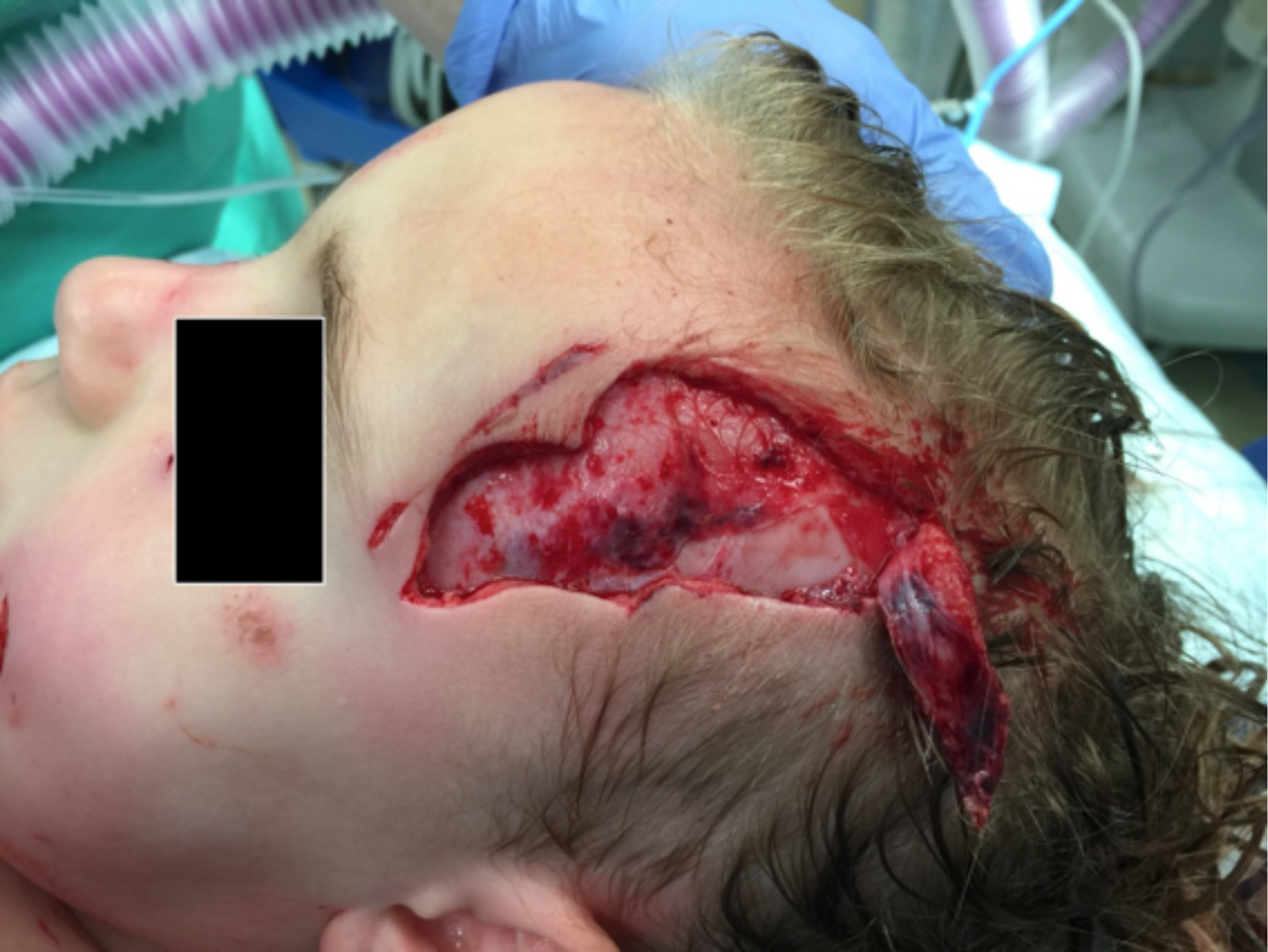 Figure 1. Scalp/forehead avulsion
The wound was then inspected for level of injury. In the forehead, the frontalis muscle was intact and no nerve injury was found. If frontal nerve injury is encountered and suitable proximal and distal nerve branches are found, nerve repair is performed. Next, meticulous closure of the muscle that has been lacerated is performed using 4-0 monocryl suture. The scalp and forehead were then closed taking care to align anatomic landmarks including the hairline of the frontal scalp. The patient did very well and had no long term morbidity.
DISCUSSION
Kaye et al published a large series of pediatric dog bite injuries. They note this is an under recognized public health concern. They treated 551 patients over a 5 year period, ages 5 months to 18 years. The incidence of injury in preschoolers (ages 2 to 5) was 24 percent. Nearly 29 percent of patients experienced bites to the face. Of note, the majority of pediatric patients experienced multiple bite wounds. Common dog breeds responsible for these bite wounds include Pit bulls, German shepherds, Rottweillers, and Akitas.
Pediatric dog bite wounds represent a significant problem encountered by the reconstructive surgeon and emergency physicians and represent a public health concern. Reconstruction principles of complex wound management are used to treat these challenging problems.
References
Kaye AE, Belz JM, Kirschner RE. Pediatric Dog Bite Injuries: A 5 year Review of the Experience at The Children’s Hospital of Philadelphia. Plast Recon Surg. 124: 551, 2009
