
Ms. Van Dine is originally from California but now resides in Utah working at the University of Utah in the Adult Reconstruction Program with Dr. Christopher Pelt. She graduated from the University of Colorado with a BA in History and obtained her master’s in physician assistant studies at the University of Utah PA program. She has been with Dr. Pelt and the University of Utah since she graduated in 2011. They have a high volume practice treating a wide variety of cases including some of the most complex primary, revision surgeries in the field. She has almost 10 years of experience assisting in the operating room, closing wounds and monitoring postoperative wound progress. When not taking care of patients she spends her time with her husband and son biking, skiing and enjoying the beauty of Utah. Ms. Van Dine is a consultant for 3M.
Van Dine_Current Dialogues in Wound Management_2020_Article_13
Introduction
Total knee arthroplasty is one of the most common surgical procedures, and in certain circumstances may lead to the increased need for revision knee surgery. Revision surgeries have a higher risk for major post-operative complications1 and, in our current healthcare climate, reducing complications is not only beneficial for patients but also for lessening the burden on our health care system and for contributing to lower overall costs. Readmissions may lead to a significant cost burden to both patients and institutions, and in some instances, can be prevented.2
Closed incision negative pressure therapy (ciNPT) is becoming more widely used by surgeons as it has shown to be beneficial in decreasing surgical site complications especially in patients with increased risk factors.3;4 Studies have shown that there can be up to a fourfold decrease in wound complications with the use ciNPT.5 We have used the PREVENA™ Incision Management System in our practice using an informal risk stratification system for the past 4 years and have seen a significant reduction in surgical site complications and positive outcomes especially in the setting of revision surgeries. The PREVENA™ System has become the standard of care for our team for most of our revision hip and knee patients who fall into our center’s informal risk stratification algorithm. The following case depicts a multiple total knee revision that was at increased risk for significant postoperative complications. Due to surgical expertise, as well as a well-planned wound and soft tissue management approach, we were able to facilitate an excellent outcome for this patient.
Case
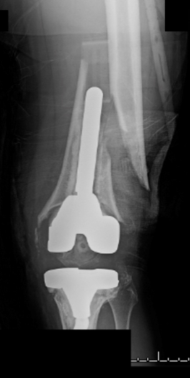
A 72-year-old otherwise healthy female with a previous total knee arthroplasty (TKA) and subsequent aseptic revision for component loosening was seen at our institution following a fall from her horse in which she sustained an open periprosthetic femoral fracture (Figure 1). The patient underwent open reduction and external fixation at an outside facility.
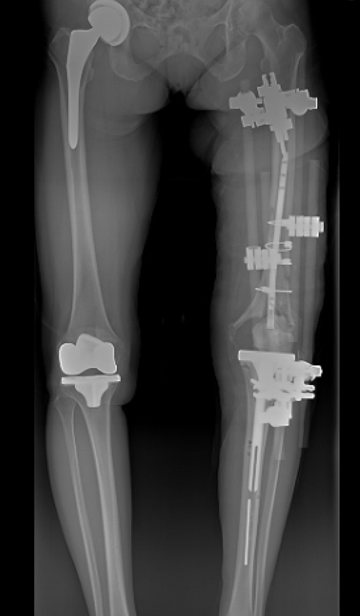
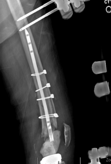
The patient had no significant past medical history but did have a surgical history of multiple left knee revisions, including a previous right total hip arthroplasty and TKA. Prior to presenting for care at our institution, the patient had undergone multiple irrigation and debridement procedures following her reduction and fixation for this knee due to concern about infection at the outside facility. Upon presentation to our clinic, external fixator pins that had been in place for 3 weeks could be seen touching the implants via radiographic imaging (Figures 2-3). The external fixator pins also displayed evidence of infection with gross purulence observed from the pin sites.
We had an extensive conversation with the patient about options to salvage her knee, but we were not overly optimistic about the likelihood of her having a well-functioning knee. We were also worried that limb amputation might be her only option. However, the patient wanted to exhaust all options for limb salvage as she was very active and was hopeful to return to her lifestyle despite this severe problem.
Pre-operative work up was positive for an infection with elevated erythrocyte sedimentation rate and C-reactive protein in conjunction with an elevated cell count on knee aspiration. The external fixator and all implants were removed and a stage I static antibiotic spacer was placed. This was the first of 3 major surgical procedures. The patient was thin with minimal soft tissue coverage over her knee, so there was concern for how the soft tissue envelope was going to react to multiple large surgeries since she had already undergone three prior knee surgeries with large incisions. The infection also necessitated a fairly large area of soft tissue excision around her old incision.
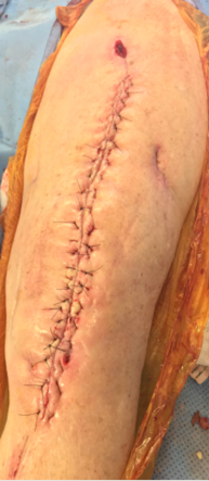
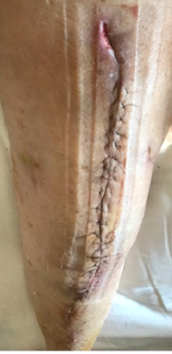
The patient’s surgery went as planned, with removal of the old hardware and placement of a static spacer. The wound closure was performed with 2-0 monocryl subcuticular interrupted suture and 3-0 nylon vertical mattress for superficial skin closure (Figure 4). The PREVENA PLUS™ Incision Management System was placed in the operating theater after final wound closure to help with wound healing, protection from external contamination, and possible drainage. Our choice to place the PREVENA PLUS™ System was consistent with our informal risk stratification algorithm as this was a revision setting with concern for the incision and surrounding soft tissue healing in a patient with low body mass index. A peripherally inserted central catheter line was placed and intravenous antibiotics were initiated per our service protocols.
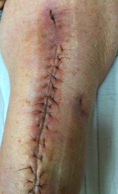
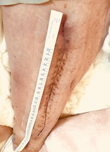
The patient returned home post operatively. Our center does not require patients to return for dressing changes but, instead, provides patients with an occlusive dressing for the home health care worker to place 5-7 days after surgery. The patient sent the care team photos of her incision that showed excellent healing and minimal postoperative swelling. She had an area superior to the surgical incision about 10 cm x 5 cm that had to be left to close by secondary intention as it was one of the more grossly infected pin sites from her external fixator, but it showed no signs of infection or delayed healing (Figures 5-6).
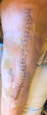
At her 3-week follow up, the patient had excellent soft tissue healing, minimal swelling and we were able to remove sutures with no healing concerns. Over the next 12 weeks, the patient had excellent healing as well with no signs or concerns for persistent infection (Figure 7).
Thirteen weeks after her stage 1 static spacer was placed, the patient’s lab results for infection had normalized and we were ready to move forward with the stage II revision. The surgeon decided to use a compress for the distal femoral replacing implant. Again, the patient’s surgery went well with no complications, and a PREVENA PLUS™ System was placed following incision closure due to the previously-mentioned risk stratification approach.
The patient returned home and was followed remotely with no postoperative wound complications. The PREVENA PLUS™ System was removed 7 days postoperatively and an occlusive dressing was placed by her home health provider. The patient initially did well but had increasing pain about 4 weeks postoperatively with an increased sensation of shifting and decreasing stability at the 8-week follow up appointment. Clinical imaging showed a fracture at the anterior cortex of the distal cut end of the bone which had allowed the spindle to bend and fracture, leaving the prosthesis incompetent. The patient was taken back to the operating room urgently to undergo distal femoral replacement. We counselled the patient about potential outcomes and the continued risk for amputation in the setting of her decreased bone stock and our concern for wound healing.
We went ahead with distal femoral replacement and the surgery went as planned. The same incision with some tissue resection was utilized further putting this wound at risk for delayed or poor healing. The incision was closed as above with 3-0 nylon for the superficial layer. The PREVENA PLUS™ System was used again, and at the 2-week follow up, the patient had excellent wound healing and no complications. The patient went on to fully heal and back to enjoying daily activities. We were able to make it through 3 complex, high-risk surgeries without surgical site complications (Figure 8).
Discussion
Total knee revisions are always more challenging and increase the chance for postoperative complications. Wound complications can be devastating in large revisions where options for further surgical treatments are limited. The PREVENA PLUS™ System has become a staple in our practice for incision management in high-risk patients. We have implemented a risk stratification protocol in our practice to guide our use; however, we believe that in the revision setting, there are few negatives to employing the PREVENA PLUS™ System.
This patient was at high risk for soft tissue failure following multiple prior revisions and use of the same incision repeatedly with the reality that any type of complication could potentially lead to further surgery and increase the likelihood of amputation. The patient went through all of her procedures with our team with no wound complications and expressed high patient satisfaction. In the end, the patient felt that she had improved cosmesis and better healing throughout her treatment with us than all of her prior surgeries. We will continue to use the PREVENA PLUS™ System in our high-risk patients as the clinical benefit warrants its use in decreasing and normalizing the risk of surgical site infections, seroma and overall wound complications.3
Patient data and photos courtesy of Christin Van Dine, PA-C, MPAS
As with any case study, the results and outcomes should not be interpreted as a guarantee or warranty of similar results. Individual results may vary depending on the patient’s circumstances and condition.
NOTE: Specific indications, contraindications, warnings, precautions, and safety information exist for PREVENA™ Therapy. Please consult the applicable PREVENA™ System Clinician Guide instructions for use prior to application. Rx only.
References
- Boddapati V, Fu MC, Mayman DJ, Su EP, Sculco PK, McLawhorn AS. Revision Total Knee Arthroplasty for Periprosthetic Joint Infection Is Associated With Increased Postoperative Morbidity and Mortality Relative to Noninfectious Revisions. J Arthroplasty 2018;33(2):521-526. doi:10.1016/j.arth.2017.09.021.
- Bosco JA, III, Karkenny AJ, Hutzler LH, Slover JD, Iorio R. Cost burden of 30-day readmissions following Medicare total hip and knee arthroplasty. J Arthroplasty 2014;29(5):903-905. doi:10.1016/j.arth.2013.11.006.
- Anatone AJ, Shah RP, Jennings EL, Geller JA, Cooper HJ. A risk-stratification algorithm to reduce superficial surgical site complications in primary hip and knee arthroplasty. Arthroplasty Today 2018;4(4):493-498.
- Newman JM, Siqueira MB, Klika AK, Molloy RM, Barsoum WK, Higuera CA. Use of Closed Incisional Negative Pressure Wound Therapy After Revision Total Hip and Knee Arthroplasty in Patients at High Risk for Infection: A Prospective, Randomized Clinical Trial. J Arthroplasty 2018.
- Matar HE, Emms N, Raut V. Effectiveness of Negative-Pressure Wound Therapy following Total Hip and Knee Replacements. J Long Term Eff Med Implants 2019;29(1):51-57. doi:10.1615/JLongTermEffMedImplants.2019030593.
© 2020 3M. All rights reserved. 3M and the other marks shown are marks and/o registered marks. Unauthorized use prohibited. PRA-PM-US-02508 (07/20).
