
Akio Kodama, M.D., Ph.D., is a Professor of Vascular Surgery at Aichi Medical University, Japan. He completed his medical training at Nagoya University and has built a distinguished career focused on chronic limb-threatening ischemia, aortic aneurysm repair, and peripheral arterial disease. A prolific researcher with over 75 peer-reviewed publications in leading journals including the European Journal of Vascular and Endovascular Surgery and Journal of Vascular Surgery, he is a board member of the Japanese Society for Vascular Surgery and a newly appointed Councilor of the World Federation of Vascular Surgery. He has received international recognition including the Takayasu Award. Dr. Kodama is a paid consultant for Solventum.
Kodama_Current-Dialogues-in-Wound-Management_2026_Article-4
Chronic limb-threatening ischemia (CLTI) represents the most advanced stage of lower extremity artery disease, a condition that is increasing in prevalence and contributes to rising global healthcare costs. CLTI is associated with significant morbidity in addition to high risks of mortality, limb amputation, and severe pain, and is accompanied by a marked reduction in health-related quality of life.1
Revascularization, such as endovascular treatment (EVT) and surgical bypass, is required to alleviate severe clinical symptoms. Recent guidelines recommend selecting the optimal revascularization plan using the PLAN framework, which an integrated three-step approach that considers patient risk estimation, limb staging, and anatomic pattern of disease.1 However, applying this approach in routine clinical practice can be challenging because of the marked heterogeneity of the CLTI population.
In Japan, patient characteristics differ substantially from those in Western countries, with approximately 50% of patients having end-stage renal failure requiring dialysis, and more than 70% having diabetes.2-4 We have previously reported clinical outcomes of patients with CLTI following revascularization in the SPINACH (Surgical reconstruction versus peripheral intervention in patients with critical limb ischemia) registry.2,5,6 Within this study, we also investigated which patients had derived greater benefit from EVT versus surgical bypass. Our analyses demonstrate that surgical revascularization is preferable in patients with wound, ischemia, foot infection (WIfI) wound grade 3 and infection grades 2 or 3. Consequently, surgical bypass is frequently performed for CLTI patients presenting with extensive ischemic wounds or severe infectious wounds. Here, I report a case of CLTI managed with revascularization and wound care using negative pressure wound therapy (NPWT).
Case
A 71-year-old woman presented with gangrene of the right fifth toe. Her medical history was notable for diabetes, end-stage renal disease on dialysis, coronary artery disease status after bypass surgery, and a prior left below-the-knee amputation. She was ambulatory with the use of a prosthesis. Initial management at the referring hospital included EVT for digital gangrene; however, worsening infection necessitated amputation of the fifth toe. Despite this intervention, the gangrene continued to worsen, and she was subsequently referred to our department. On presentation at our hospital, she had extensive dry gangrene involving the forefoot (Figure 1).

Non-invasive vascular assessment showed a markedly reduced ankle-brachial pressure index (ABI) of 0.60 and a skin perfusion pressure (SPP) of 12 mmHg. Laboratory findings on admission showed leukocytosis (white blood cell count of 12,600) and elevated C-reactive protein levels (15.89 mg/dl). Though, other signs of infection (fever, fatigue, and warmth at wound site, or purulent wound exudate) were absent. Her plain radiographs revealed severe arterial calcification (Figure 2).

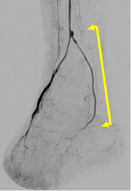
On the day following transfer, we performed a bypass from the below-knee popliteal artery to the dorsalis pedis artery (Figure 3), together with a trans-metatarsal amputation. Iodine-based ointments and standard wound care dressings were used for wound care with daily dressing changes. By postoperative day 23, ABI and SPP values had improved; however, the plantar wound had worsened and showed no evidence of granulation tissue formation (Figure 4).


To enhance plantar perfusion, we performed an additional bypass from the previous graft to the medial plantar artery on postoperative day 24 (Figure 5). Following this procedure, plantar SPP values improved. Solventum™ V.A.C.® Therapy was initiated using Solventum™ V.A.C.® Granufoam™ Dressing with continuous pressure at -125 mmHg. Dressings were changed every 3 days. V.A.C.® Therapy was discontinued after 4 weeks when the wound bed was covered with healthy granulation tissue. A split-thickness skin graft procedure was performed. The donor site on the patient’s thigh and the skin graft over the wound were covered with standard post-surgical dressings and iodine-based ointment. The dressing changes occurred daily (Figure 6). Wound closure was successfully achieved through the combination of vascular surgery to restore blood flow to the foot, standard wound care dressings, V.A.C.® Therapy, and skin grafting.

References
- Conte MS, Bradbury AW, Kolh P, et al. Global vascular guidelines on the management of chronic limb-threatening ischemia. J Vasc Surg. 2019;69(6S):3S-125S.e40. doi:10.1016/j.jvs.2019.02.016
- Iida O, Takahara M, Soga Y, et al. Three-year outcomes of surgical versus endovascular revascularization for critical limb ischemia: the SPINACH study (Surgical reconstruction versus peripheral intervention in patients with critical limb ischemia). Circ Cardiovasc Interv. 2017;10(12):e005531. doi:10.1161/CIRCINTERVENTIONS.117.005531.
- Farber A, Menard MT, Conte MS, et al. Surgery or Endovascular Therapy for Chronic Limb-Threatening Ischemia. N Engl J Med. 2022;387(25):2305-2316. doi:10.1056/NEJMoa2207899
- Bradbury AW, Moakes CA, Popplewell M, et al. A vein bypass first versus a best endovascular treatment first revascularisation strategy for patients with chronic limb threatening ischaemia who required an infra-popliteal, with or without an additional more proximal infra-inguinal revascularisation procedure to restore limb perfusion (BASIL-2): an open-label, randomised, multicentre, phase 3 trial. Lancet. 2023;401(10390):1798-1809. doi:10.1016/S0140-6736(23)00462-2.
- Kodama A, Takahara M, Iida O, et al. Health related quality of life over time after revascularisation in patients with chronic limb threatening ischaemia. Eur J Vasc Endovasc Surg. 2021;62(5):777-785. doi:10.1016/j.ejvs.2021.06.037
- Kodama A, Takahara M, Iida O, et al. Ambulatory status over time after revascularization in patients with chronic limb-threatening ischemia. J Atheroscler Thromb. 2022;29(6):866-880. doi:10.5551/jat.62892
Patient data and images courtesy of Akio Kodama, MD, PhD.
NOTE: Specific indications, contraindications, warnings, precautions and safety information exist for these products and therapies. Please consult a clinician and product instructions for use prior to application. Rx only.
As with any case study, the results should not be interpreted as a guarantee or warranty of comparable results. Individual results may vary depending on the patient’s circumstances and condition.