
Dr. A.J. Applewhite received his undergraduate degree from the University of Texas in Austin, his medical degree from the University of Texas Medical School in Houston and completed his Family Medicine Residency through UTMB. He is board certified in family medicine as well as Undersea and Hyperbaric Medicine. He is a Certified Wound Specialist Physician, Fellow of the American Professional Wound Care Association and the current President of the Gulf Coast Chapter of the Undersea and Hyperbaric Medical Society. Dr. Applewhite is the Medical Director of the Comprehensive Wound Center at Baylor University Medical Center and an Associate Clinical Professor at Texas A & M Medical School.
Applewhite_Current Dialogues in Wound Management_2019_Volume 5_Issue 1
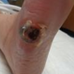 Figure 1. Wound upon initial presentation
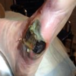 Figure 2. Wound 7 days after initial presentation

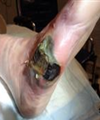
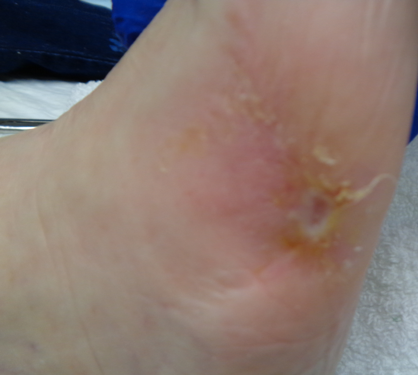
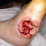 Figure 3. Wound after surgical debridement (19 days post initial presentation)
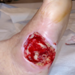 Figure 4. Wound after 3 days of V.A.C. VERAFLO™ Therapy
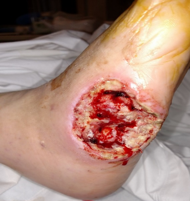

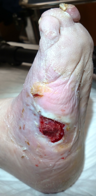
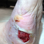 Figure 5. Wound at post-operative day 30
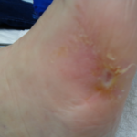 Figure 6 Wound fully healed at approximately 90 days after surgery.
INTRODUCTION
As our population ages and obesity increases, diabetes has become a major medical concern. In fact, the incidence and prevalence of diabetes doubled from 1990 to 2012.1 As a result of its many microvascular and macrovascular complications including coronary artery disease, end-stage renal disease, stroke, retinopathy, and foot ulcers, the total expenditures on these complications account for nearly 14% of the total health care expenditure in the United States.2 While not garnering the national attention of heart disease, kidney disease or stroke, diabetic foot ulcers (DFUs) are a potentially deadly and costly complication of diabetes. Comprehensive wound management involving a multidisciplinary team is required to heal DFUs and avoid amputations,3 which may include specialized dressings, debridement of necrotic tissue, management of blood sugars and management of infection, offloading pressure with a total contact cast, hyperbaric oxygen therapy, and revascularization. Approximately 85% of all non-traumatic amputations are preceded by an ulcer.4 Nearly half of the patients who receive a major leg amputation will die within 5 years,5 and around 55% will undergo a contralateral amputation within 2-3 years.6 Therefore, it is imperative to aggressively treat DFUs in order to prevent amputations. We present, here, the case of a complex situation which illustrates how a concerted effort by members of a multidisciplinary team contributed to limb salvage of a woman’s leg.
CASE STUDY
A 68-year-old female presented to the wound center with a right lateral diabetic foot ulcer which had appeared approximately one month earlier. Her past medical history included type II diabetes mellitus (DM), congestive heart failure, hypertension, hypothyroidism, hyperlipidemia, peripheral arterial disease (PAD), breast cancer, cervical cancer, a left below-the-knee amputation (BKA) and a right 1st and 5th toe amputation. She was a former smoker but had quit six years prior to the left BKA. The patient ambulated well with her left leg prosthesis, and the wound was a relatively small full thickness ulcer with 100% eschar (Figure 1). The wound was dry and black and had no evidence of acute infection. The patient’s distal pulses were weak, and her ankle-brachial pressure index (ABI) was 0.68. She had been evaluated at another facility, undergone an unsuccessful angioplasty and was told she needed a right below-the-knee amputation. Not wanting to become a double amputee, the patient sought a second opinion. She stated that her blood sugar was controlled and denied having any fever, chills, nausea, vomiting or significant pain.
After a thorough evaluation of her medical history, physical exam and ABI by the wound care specialist, it was determined that the main issue was the PAD causing a diabetic foot ulcer (DFU) with dry gangrene. As it appeared stable, an appointment was made with our facility’s vascular surgery group within the week. An antimicrobial dressing was applied to the wound, and she was instructed to offload the foot and minimize walking using a Darco Wedge and rolling walker. Unfortunately, the patient was unable to be seen at the vascular surgeon’s office due to financial issues. The patient became depressed and resigned herself to having another BKA. The patient started walking without regard to offloading the wound. She returned to the wound center exactly one week after her initial presentation with a significantly worsening wound, which had turned from dry gangrene to wet gangrene and had become larger, boggy and malodorous (Figure 2). In addition, the patient reported increased pain, nausea and chills. She was immediately admitted to the hospital and underwent a bedside debridement by a podiatry specialist, and intravenous (IV) antibiotics were initiated by the infectious disease specialist. An MRI revealed osteomyelitis. The patient’s blood sugars were uncontrolled, with an HgbA1C on admission of 11.2 mmol/L, and were subsequently managed by the internal medicine team and she was started on insulin.
A vascular surgery specialist performed an angiogram which revealed significant superior femoral artery occlusion which required a distal bypass in order to restore adequate circulation to the foot. With her history of heart failure, the patient required cardiac clearance prior to any major surgery. A heart catheterization by the cardiologist revealed significant three-vessel disease, and she received a heart stent. Her ejection fraction was noted to be 22%. Two days later, the patient received a right femoral popliteal bypass. Surgical debridement was performed by a podiatrist but was shortened due to cardiac issues noted during surgery, leaving the wound with a moderate amount of devitalized tissue (Figure 3). V.A.C. VERAFLO™ Therapy using the V.A.C. VERAFLO™ Dressing was initiated by the wound care specialist (Figure 4). Normal saline was instilled into the wound bed with a 10-minute dwell time followed by 3 hours of negative pressure at -125 mmHg. Dressing changes occurred every 3 days. The patient was deemed not cleared for any further surgery by cardiology. With the patient stabilized and no further surgery planned, discharge planning was discussed across the care team to manage any potential excess hospitalization costs that may not have been covered by the patient’s insurance plan. Accordingly, at the 3-day dressing change, the wound had significantly improved, and the patient was approved for discharge to a skilled nursing facility (SNF) for continued IV antibiotics, wound care with V.A.C.® Therapy, and rehabilitation.
Approximately one month later, the patient was discharged from the SNF after receiving a total of 6 weeks of IV antibiotics and was seen in the wound center. While in the facility, the patient participated in physical therapy, but this was limited secondary to the V.A.C.® Therapy, and non-weight bearing status of the right foot and the patient had become quite deconditioned. The wound had improved but was still significant (Figure 5). Hyperbaric oxygen therapy (HBOT) was also added to the patient’s care plan. As the wound improved, approximately two weeks after HBOT was initiated, V.A.C.® Therapy was discontinued, and total contact casting (TCC) with PROMOGRAN PRISMA™ Matrix was applied, thus allowing the patient to ambulate with her prosthesis and improve her conditioning. Approximately 8 weeks later, after 30 HBOT treatments and 9 cast placements, the wound healed (Figure 6) and the patient was able to walk out of the wound center with her leg intact. She was able to maintain an independent life and perform all the usual activities of daily living.
DISCUSSION
This case demonstrates the complexities involved in treating DFUs and the coordinated teamwork required to manage the multiple comorbidities which complicate the care of these patients, which may be further compounded by extrinsic factors and social situations. For this patient, her social situation created a downward spiral which nearly led to an amputation. Her journey took her through the entire continuum of care from the outpatient clinic setting to the acute care inpatient setting to the SNF and back to the outpatient clinic. Her cardiac status prevented aggressive debridement, necessitating a highly specialized wound dressing and without a distal bypass, her circulation would not have been adequate to heal the wound. In the end, this patient required an “all hands-on-deck” approach in which every member of her care team contributed to limb salvage. If anything had gone wrong, it would have resulted in a BKA, which would have turned our patient into a double amputee effectively unable to care for herself and entirely reliant on others. In addition, this could have led to more depression, decreased quality of life and most likely, placement in a nursing home. It is important to understand that we are not just treating the wound, but we are treating the patient with the wound.
References
1. Geiss LS, Wang J, Cheng YJ, et al. Prevalence and Incidence trends for diagnosed diabetes among adults aged 20-79 Years, United States, 1980-2012. JAMA. 2014;312(12):1218-1226
2.Lin P, Phillips T. Ulcers. In: Blognia J, Jorrizo, J, Rapini R. Dermatology. Vol 2. Philadelphia, PA: Elsevier; 2003:1631-1649.
3.Snyder RJ, Kirsner RS, Warriner RA III, et al. Consensus Recommendations on Advancing the Standard of Care for treating Neuropathic Foot Ulcers in Patients with Diabetes. Ostomy Wound Management. 2010;56(Suppl 4):S1-S24
4.Alexiadou K, Doupis J. Management of diabetic foot ulcers. Diabetes Ther. 2012;3(1):4.
5.Bild D, Selby J, Sinnock P, et al., Lower-extremity amputation in people with diabetes. Epidemiology and prevention. Diabetes Care. 1989; 12(1): 24-31
6.Most RS, Sinnock P. The epidemiology of lower extremity amputations in diabetic individuals. Diabetes Care. 1983; 6(1): 87-91
Photos and patient information courtesy of Andrew J. Applewhite, MD, CWS, Baylor University Medical Center, Comprehensive Wound Center, Dallas, TX.
As with any case study, the results and outcomes should not be interpreted as a guarantee or warranty of similar results. Individual results may vary depending on the patient’s circumstances and condition.
NOTE: Specific indications, contraindications, warnings, precautions and safety information may exist for Systagenix and KCI (Acelity companies) products. Please consult a healthcare provider and product instructions for use prior to application. Rx only.
