
Jayesh B. Shah, MD, CWSP, UHM, is the President of South Texas Wound Associates, PA, San Antonio, Texas, where he provides clinical wound care services in San Antonio and the surrounding communities. He is also the President of TIMEO2 Healing Concepts, LLC in San Antonio, Texas, which
provides consulting and education services in wound care and hyperbaric medicine both nationally and internationally.His degrees include MBBS from M. S. University, India and MD in Internal Medicine from St. Luke’s Roosevelt Hospital, Columbia University, New York. He is Board Certified in Internal Medicine, Board Certified in Undersea and Hyperbaric Medicine, Certified in Wound Management, Certified in Hyperbaric Medicine, Past Chair of American College of Clinical Wound Specialists and Past Vice President of American College of Hyperbaric Medicine. He has been the recipient of the Enterpreuner of the year award by the Alamo Asia Chamber of Commerce (2016); the Paul James Sheffield Education Award for Lifetime Dedication to Education in the field of Undersea and Hyperbaric Medicine (2014); Jefferson C. Davis Memorial Award for Excellence in Clinical Hyperbaric Medicine (2007 and 2011); Carolyn Sue Award (2009); Young Scientist/Medical Doctor Award (2008); Community Service and Leadership award by Alamo Asian American Chamber of Commerce (2008). He has published 3 books- “Wound Care Certification Study Guide” in 2011 with its second edition 2016, Textbook of Clinical Wound Medicine: A Evidence Based Approach in press for release in 2017. He has authored over 40 chapters on various wound topics in 4 books in addition to 30+ scientific articles in wound care and hyperbaric medicine. As an Assistant Editor of the Journal of ACCWS, he regularly writes a column on certification exam in wound care.
Shah_Advanced Wound Dressings Today_Advanced Wound Dressings Today_2017_Spring Supplement
INTRODUCTION
Management of infection and biofilm are necessary for wound bed preparation.1 Silver has a broad spectrum of antimicrobial activity including activity against MRSA and VRE.2 Although use of silver has been described in history since the ancient times, 3 it really became more popular after 1970’s when silver was used in treatment of burns using either 0.5% silver nitrate solution or topical cream like silver sulfadiazine.4 Recently, there is a newer class of silver dressings available in a variety of forms like transparent dressings, gauze, island dressings, foams, and absorptive filler. Silver dressings are also available with various combinations like collagen, hydrofiber, alginates, foam, honey and hydrogels.4 These dressings are designed to provide the antimicrobial activity of topical silver in a more convenient application.4 These silver dressings differ considerably in the amount of silver ions and their physical and chemical properties.2, 4
MECHANISM OF ACTION
Silver ions bind to the bacterial wall at multiple sites, causing membrane damage and cytoplasm leakage, which makes some silver dressings bactericidal.4 Silver ions also bind to the cell proteins, causing cell death and destroying cell DNA. 4
NEWER SILVER DRESSINGS AND EVIDENCE
Newer silver dressings have silver in form of complexes of silver salts or nanocrystalline silver metal. It may be found as element Ag or ion Ag+.4, 5 Sibald et al studied the use of nanocrystalline silver primary dressing on chronic venous stasis ulcer patients who failed multilayer compression therapy.Study results found that silver dressings reduces bacterial counts, increases lymphocyte count and improves healing rates4, 6 in a statistically significant way. Castellano et al. compared eight silver containing dressings and concluded that dressings with higher concentration of silver ions may be more appropriate for wounds that contain 105 organisms.4, 7 Similarly, Gago et al compared healing of venous ulcers with silver dressing and concluded that patients who had higher concentration of silver in dressing showed reduced healing time and quicker resolution of infection.4, 8 While Parson’s et al found that there was no correlation between antibacterial effect and silver content of dressings.2
CASE STUDY
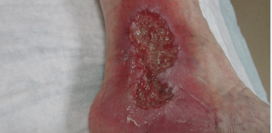
In a recent case, a 60 year old female who works as a cashier and stands on her feet all the time presented to my office. The patient had a history of hypertension, a history of venous stasis and a history of ulcers in the past which were treated with compression therapy. The patient was not wearing stockings and she developed ulcer on left ankle which started from rubbing of shoes. She initially presented with a blister which progressively got worse. So she was sent to the wound care center for a complete comprehensive evaluation. Upon deeper examination, the patient had normal arterial doppler with triphasic blood flow. On admission we started the patient on calcium alginate with silver and conducted debridement.Her ABI Right measured 1.02 with the left being non compressible. So, we obtained a wound culture, started the patient on Cefdinir 300mg 1 tab PO BID X10 days for one week. Ultimately, the wound got worse so we then started the patient on Dakin’s solution 0.25% daily for 1 week (see Figure 1).
Figure 1: A 6.6 cm X 2.7 cm X 0.4 cm wound
At the two week mark, we started the patient on silver powder mixed with iodine-based gel with a compression dressing 3 times per week. At the one month mark, the wound was still worse with a wound size of 6.8cm x 3 cm x 0.3cm with 100% soft necrotic tissue and purulent drainage.The patient was started on 0.25% Dakin’s solution with daily dressing changes. In addition the patient was admitted to hospital where a culture was run for multidrug resistant pseudomonas and the patient was also started on IV Merrem® for four weeks (see Figure 3). At the two month mark, the patient was discharged from a rehab facility after 4 weeks of IV Merrem®. The wound size was now smaller at 5cm x 1.5cm

Figure 2: Wound has increased in size to 6.9 cm X 2.6cm X 0.2cm with continued purulent drainage.

Figure 3: Wound at the one month mark with 100% soft necrotic tissue

Figure 4: Wound at 8 week mark
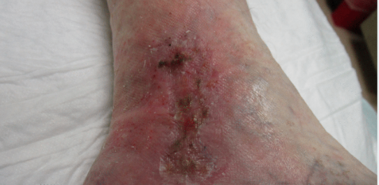
Figure 5: Wound at ~12 week mark
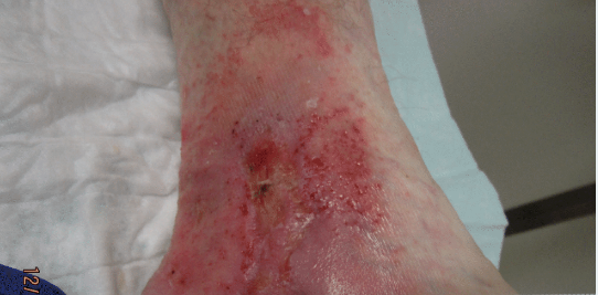
Figure 6: Wound at ~ 3.5 month mark

Figure 7: Wound at discharge (~4.5 month mark)
x 0.2cm with 30% of the wound area having commenced epithelization. In addition, there was 50% granulation tissue and minimal slough. We then started the patient on Collagen Silver Dressing and a compression bandage (see Figure 4). At the approximate three month mark since presentation, we continued the patient on Collagen-Silver and a compression bandage. The wound continued to epithelialize (~ 80%) and the wound size had now diminished to 1cm x 0.3cm x 0.1cm (see Figure 5)
At the three and a half month mark, the wound has achieved significant epithelialization. The patient was started on amylactin lotion and continued use of a compression bandage. The wound size is now measured at 0.8cm X 0.3cm X 0.1cm with 80 -90% epithealialization (see Figure 6). About a week after this visit, the patient’s wound had epithelialized 100% and now measured 0.1cm x 0.1cm x 0.1cm and was managed with amylactin lotion and a compression bandage. After a few more weeks of treatment and follow up, the patient was discharged from our care with a class II compression stocking (see Figure 7).
CONCLUSION
Choice of wound dressing should be based on the wound type and clinical parameters such as exudate handling effects and presence of biofilm or critical colonization. 4 Dry wounds should receive moist dressings and wet wounds should receive absorbent dressings. Based on clinical studies, general recommendation can be made that dressings with higher silver can be used in refractory wound with recurrent biofilm and in clinically infected wounds.4, 7 Clinically, 10 minute 0.25% acetic acid or 0.25% Dakin’s solution soaks are recommended to treat biofilms prior to the application of silver based dressings. Dressing with lower concentration of silver or combination of collagen and silver could be considered in wounds that are stalling or suspected of being colonized or critically colonized.4, 7, 8 Studies are not able to recommend specific length of time for use of silver dressings but it is prudent to not use silver dressings for more than 9 -12 weeks. Long term use of silver may be associated with systemic toxicity though Sibald et al found that there was only a slight increase in blood silver concentration indicating that nanocrystalline silver dressings are not systemically toxic 6 while silver sulfadiazine does contain fast release silver which was easily absorbed by tissues and was associated with systemic toxicity.4, 9
References
1.Shah JB, Correction of hypoxia a critical element of wound bed preparation guidelines: TIMEO2 Principle of wound bed preparation.J Am Col Certif Wound Spec. 2011 Oct 9;3(2):26-32. doi: 10.1016/j.jcws.2011.09.001.
2.Parson’s et al, Silver Antimicrobial Dressings in Wound Management: A comparison of Antibacterial, Physical, and Chemical characteristics: Wounds 2005:17(8): 222-232.
3.Shah JB, History of Wound care, J Am Col Certif Wound Spec. 2011 Sep;3(3):65-6. doi: 10.1016/j.jcws.2012.04.002.
4.Rose L. Hamm, Antibacterial Dressing, Advances in wound care by Chandan K. Sen, Volume 1, 2010, 148 -154 Volume 1.Publisher Mary Ann Liebert, Inc. Publications.
5.Rodeheaver GT and Ratiff CR; Wound cleansing, wound irrigation, wound disinfection, in Chronic Wound Care: A Clinical Source Book for Health-Care Professionals, 4th Edition, edited by Krasner DL, Rodeheaver GT, and Sibbald RG, Malvern PA. HMP communications: 2007, pp. 331-342.
6.Sibald RG et al: Bacteriology, Inflammation, and healing: a study of nanocrystalline silver dressings in chronic venous leg ulcers. Adv. Skin Wound Care 2007:20:549.
7.Castellano et al: Comparative evaluation of silver-containing antimicrobial dressings and drug. Int. Wound J. 2007:4:114.
8.Gago et al: A comparison of three silver containing dressings in treatment of infected chronic wounds. Wounds 2008:20:273.
9.Medscape: Silver sulfadiazine top, Available at www.medscape.com/druginfor/dosage?cid=med&drugid=13530&drugname=Silver+S, accessed Jan.15, 2017
REFERENCES
1. Cowan, Linda, et al. Caution: when combining topical wound treatments, more is not always better. Wound Practice & Research: Journal of the Australian Wound Management Association 19.2 (2011): 60.
2. Armstrong D, Bates-Jensen B, Bohn G, et al. Expert recommendations for the use of hypochlorous solution: science and clinical application. Wounds. 2015.
3. Scanlon, E., and Stubbs, N. To use or not to use? The debate on the use of antiseptics in wound care. British journal of community nusring 7. Sup2 (2002): 8-20.
4. Atiyeh, B. S., Dibo, S. A., & Hayek, S. N. (2009). Wound cleansing, topical antiseptics and wound cleansing, topical antiseptics and wound healing. International Wound Journal, 6(6), 420-430.
5. Drosou A, Falabella A, Kirsner RS. Antiseptics on Wounds: An Area of Controversy. Wounds. 2003; 15(5):149-166.
6. Mulder GD, Cavorsi JP, Lee DK. Antimicrobial Agents in Wound Care. Wounds. 2007;19(7):173-182.
7. McDonnell G, Russell AD. Antiseptics and disinfectants: activity, action, and resistance. Clin Microbiol Rev.1999; 12(1):147-179.
8. Senior N. Some observations on the formulation and properties of chlorhexidine. J Soc Cosmet Chem. 24(4): 259-278.
9. Basrani BR, Manek S, Sodhi R, Fillery E, Manzur A. Interaction between sodium hypochlorite and chlorhexidine gluconate. J Endod. 2007; 33(8): 966-969.
10. Yasuda K, Ohmizo C, Katsu T. Potassium and tetraphenylphosphonium ion-selective electrodes for monitoring changes in the permeability of bacterial outer and cytoplasmic membranes. J Microbiol Methods. 2003;54(1):111-115.
11. Jovanovic A, Ermis R, Mewaldt RS, Shi L, Carson D. The Influence of Metal Salts, Surfactants, and Wound Care Products on Enzymatic Activity of Collagenase, the Wound Debriding Enzyme. Wounds. 24(9):242–253.
12. Safety and effectiveness of consumer antiseptics; topical antimicrobial drug products for over-the-counter human use; proposed amendment of the tentative final monograph; reopening of administrative record; proposed rule. Fed Regist 78:76444–76478. http://www.gpo.gov/fdsys/pkg/FR-2013-12-17/pdf/2013-29814.pdf
13. Khan M, Naqvi AH. Antiseptics, iodine, povidone iodine and traumatic wound cleansing. J Tissue Viability. 2006; 16(4):6-10.
14. Smith RG. Critical discussion of the use of antiseptics in acute traumatic wounds. J Am Podiatr Med Assoc. 2005; 95(2):148-153.
15. Schmitz G. Iodine oxidation by hydrogen peroxide and Bray–Liebhafsky oscillating reaction: effect of the temperature. Phys Chem Chem Phys. 13(15): 2011: 7102-7111. doi: 10.1039/C1CP00006C
16. Sakarya S, Gunay N, Karakulak M, Ozturk B, Ertugrul B. Hypochlorous Acid: an ideal wound care agent with powerful microbicidal, antibiofilm, and wound healing potency. Wounds. 2014; 26(12):342-350.
17. Thorn RM, Lee SW, Robinson GM, Greenman J, Reynolds DM. Electrochemically activated solutions: evidence for antimicrobial efficacy and applications in healthcare environments. Eur J Clin Microbiol Infect Dis. 2014; 31(5):641–653.
18. Edwards K. New Twist on an Old Favorite: Gentian Violet and Methylene Blue Antibacterial Foams. Adv Wound Care(New Rochelle). 2016; 5(1): 11–18.
19. Hydrofera Blue Classic Antibacterial Foam Dressing; SDS HB2214/HB4414-00 Safety [Online]; Hollister: Libertyville, IL, April 13, 2016.http://www.hollister.com/~/media/files/pdfs%E2%80%93for%E2%80%93download/sds/sds%E2%80%93hydrofera%E2%80%93blue%E2%80%93classic.pdf?la=en (accessed April 15, 2017)
20. Chao C, Runde D. Tap Water vs. Sterile Saline for Wound Irrigation. Am Fam Physician. 2015; 92(3). http://www.thennt.com/nnt/tap-water-for-wound-irrigation/. Accessed February 10, 2017
21. Kim P, Attinger C, Crist B, et al. Negative Pressure Wound Therapy with Instillation: Review of Evidence and Recommendations. Wounds. 2015; 27(12):S2-S19.
22. Wolvos T. Wound Instillation — The Next Step in Negative Pressure Wound Therapy. Lessons Learned from Initial Experiences. Ostomy Wound Manage. 2004; 50(11):56-66.
