
Liz McElroy is a Nurse Practitioner in Wound, Ostomy, and Continence Care at the Reading Health System in West Reading, PA. She graduated from Gwynedd Mercy University with her MSN in Adult Nurse Practitioner in 2009, became a Certified Wound Specialist (CWS) in 2010, and certified in Wound, Ostomy, and Continence (CWOCN) in 2012. She serves as an in-patient wound, ostomy, and continence specialist for a 647-bed Level II Trauma Center. She focuses on care of the perioperative, post-operative, cardiac and critical care patients, with an interest in nursing and physician education and V.A.C. VERAFLO™ Therapy. She also has two years experience in an outpatient wound care center prior to her acute care position.
McElroy_Current Dialogues in Wound Management_2016_Volume 2_Issue 3
NOTE: As with any case study, the results and outcomes should not be interpreted as a guarantee or warranty of similar results. Individual results may vary depending on the patient’s circumstances and condition.
The first time I encountered lymphedema was 12 years ago while in nursing school for my BSN. I was on a home care visit with my instructor visiting a home bound patient who lived in a trailer at the back of a mini golf course. I remembered looking at the woman’s malodorous, weeping, huge legs and asking my instructor, “Is that elephantiasis?” Since then I have seen and learned so much about lymphedema, but still struggle with the shared challenges of effectively treating such patients. The entire medical team struggles to find quality of life for these patients and prevent the life altering complications that come with lymphedema and massive localized lymphedema (MLL).
Massive Localized Lymphedema was first described in 1998 in patients with morbid obesity, and its incidence has since been rising due to the increased prevalence of morbid obesity.² Chopra, et al found that 48.5% of MLL was found to be in the thigh region.³ MLL can provide significant disturbances in ability to function and quality of life. Additionally, there can be a dearth of resources for patients to obtain or afford proper interventions for this diagnosis.
I met the patient in August of 2015. She had been diagnosed with lymphedema eight years earlier after suffering for years with edema and thigh lobules, limiting her functional ability, but she was still active outside her home. She was referred for manual lymphatic decompressive therapy and compression wraps, which helped reduce her leg size, but did not stop the progression of her lymphedema, which ultimately resulted in MLL.
She had been a patient at my healthcare system’s wound care center for several months with a wound on a medial thigh lobule that was literally dragging on the ground. This lobule limited her ambulation and caused her pain. We are fortunate to have a surgeon willing to surgically excise MLL when the complications are causing the patient’s functional status decline. This is a technically difficult surgery with high risk of complications. On August 19, 2015 she underwent an excision of a MLL. She shared with me this picture of her before her surgery (Figure 1).
 Figure 1. MLL of the right thigh prior to excision
She developed a surgical dehiscence and wound infection at the excision site on POD #10, returning to the OR for washout. The surgical team treated the wound with 0.25% Acetic Acid packing changes BID. She had a lot of pain with the dressing changes, requiring IV Dilaudid, and it required at least two staff members to perform. On POD #15 she returned to the OR for a second washout, and the wound was loosely closed with sutures. The plastic surgery team asked the in-patient WOCN team to evaluate on POD #21. At this time, we removed the sutures and applied V.A.C. VERAFLO™ Therapy (Figure 2).
The surgical team wanted to continue use of Acetic Acid, so we utilized the 0.25% Acetic Acid for our instillation solution. VERAFLO™ Therapy was connected, instilling 100mL of Acetic Acid for a five minute dwell every 6 hours. At the next dressing change, we transitioned to NSS as the instillation
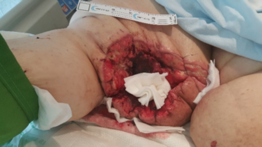 Figure 2. POD #21, V.A.C. VERAFLO™ Therapy applied. Wound measured 34.5 x 15 x 14.5cm
solution, only requiring 80 ml. One week after VERAFLO™ Therapy was initiated, the patient was ready to be discharged to the acute rehab hospital. We transitioned her to V.A.C.® Therapy (Figure 3). In the one week we had VERAFLO™ Therapy on, her wound volume decreased from 7503 cm³ to 3059 cm.³ Her pain was greatly decreased, and she now required IV narcotics for dressing changes only, not around the clock.
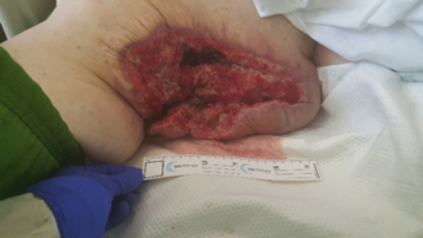 Figure 3. One week after V.A.C. VERAFLO™ Therapy wound measurements 23 x 14 x 9.5 cm
We continued to follow the patient weekly for her V.A.C.® Therapy, coordinating with the plastic surgery team. She continued to show great improvement, and after three weeks of V.A.C.® Therapy, she underwent a STSG to cover the deficit. V.A.C.® Therapy was used over the STSG, and she was discharged from the rehab one week later with closure.
I still keep in touch with the patient, and she continues to struggle with her lymphedema and thigh lobules, but is able to return to work and live independently in the community. A lesson we learned as a medical team was that the patient may have also benefitted from the use of an intact incisional NPWT, such as the PREVENA™ Incision Management System. As a profession, we also must do a better job at recognizing lymphedema early and intervening with weight loss coaching, compression therapy and referrals to lymphedema specialists early on in the disease process to decrease and prevent further complications. As new research emerges, it would be a priority to focus on prevention and treatment of MLL with
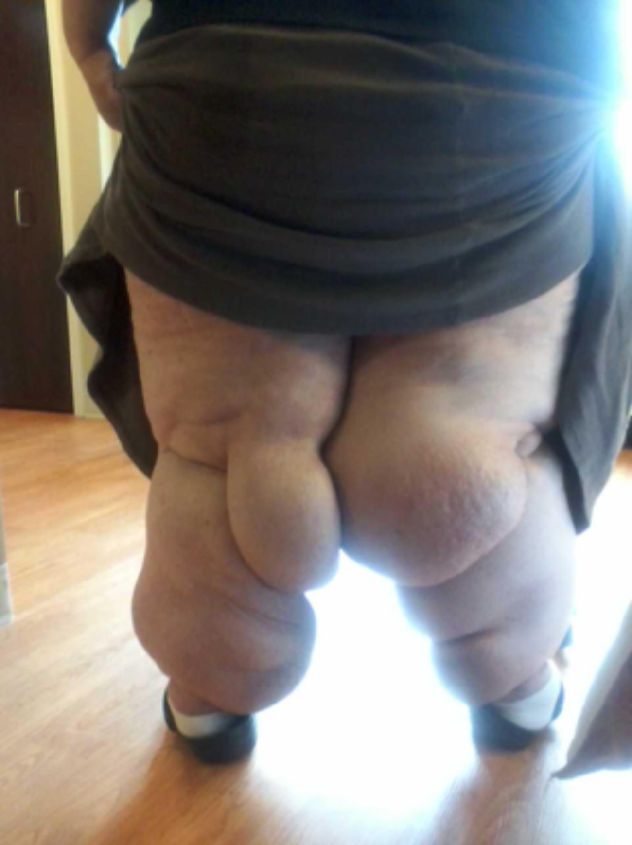 Figure 4. MLL after excision and wounds healed
a team approach including physical therapists, nurses, physicians and surgeons.
References
1.Fife C. Massive localized lymphedema, a disease unique to the morbidly obese: a case study. Ostomy Wound Management, 2014;60(1):30-35.
2.Furrukh J, et al. The diagnostic and surgical challenges of massive localized lymphedema. Am J Surg. 2015;209 (3):584-587.
3.Chopra K, Tadisina KK, Brewer M, et al. Massive localized lymphedema revisited: a quickly risking complication of the obesity epidemic. Ann Plast Surg. 2015;74(1):126-132.
4. Farshid G, Weiss S. Massive localized lymphedema in the morbidly obese: a histologically distinct reactive lesion simulating liposarcoma. Am J Surgical Pathology. 1998; 22: 1277-1283.
5.McCrystal D, O’Loughlin B. Massive localized lymphedema of the thigh. ANZ J Surg. 2007;77:91-92.
