
Kara Couch is a family nurse practitioner and Certified Wound Specialist, who has been working in wound care for the past 12 years. As a nurse scientist, she is an investigator on several clinical trials related to wound healing. Kara also serves as a member of the Guidelines Committee for the Association for the Advancement of Wound Care and has authored or co-authored numerous articles and chapters on wound healing. She also lectures nationally on wound care and wound healing topics.
Couch_Current Dialogues in Wound Management_2015_Volume 1_Issue 4
Donor sites are used to harvest an autograft or flap to close an open wound. The troublesome aspect of donor sites is that a wound is made to heal another wound. Surgeons ensure the recipient site is optimized to receive the graft. When the donor site wound does not heal as expected within the first few weeks, a wound care challenge is created.
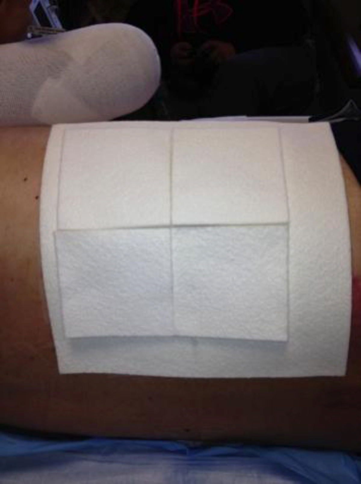
A typical donor site location for a split-thickness skin graft is the anterolateral thigh. Other sites can include the buttocks or flanks, but those sites create some challenges with postoperative pain control, as patients are resting on the sites. A partial-thickness wound is created by the use of a dermatome, and the harvested skin is then secured to the recipient site. The donor site wound is typically highly exudative, especially within the first 48 hours, and the dressing often becomes saturated.
CHOOSING A DRESSING
Numerous randomized controlled and comparative effectiveness trials, as well as several meta-analyses, evaluate donor site dressing selection.1-8 These studies show there is no one “ideal” donor site dressing to heal the wound.1 Therefore, there is broad variability to how the donor site is dressed immediately postoperatively, and dressing selection is often provider dependent. The goals of choosing a dressing for the donor site wound are to manage exudate, control pain, provide occlusion, provide impermeability to bacteria, remain intact for approximately 1 week, and minimize scarring. Other factors in choosing a dressing include issues such as cost, ease of application, pain control, and need for reapplication. Many studies have compared various dressings, but none has generated high-level evidence, and all systematic reviews have been unable to reach a consensus on dressings due to the poor quality of the studies.
Traditionally, dressings such as polyurethane films and paraffin gauze have been used over donor sites. More recently, polyurethane foams, carboxymethlycellulose (CMC) hydrofiber dressings, calcium alginates, and composite dressings have been studied. The goal again is to wick away the exudate into the dressing to allow rapid re-epithelialization. Among current dressings, alginates and polyurethane films are commonly used and studied.
MANAGING DRESSINGS
There are caveats to use of several more common dressings. Polyurethane films are applied over the donor site wound in the operating room, and the donor site exudes a moderate-to-copious amount of exudate that creates a bulge in the dressing; exudate can leak onto the patient’s clothing or disrupt the seal of the film. An easy fix for this issue is to score the film dressing and then apply an absorptive secondary dressing over the film to capture the exudate.2 Another common dressingis paraffin gauze over the site. Ideally, the paraffin gauze dressing is left in place to dry out the wound; the dressing can then be easily removed with a healed donor site underneath. Patient education on this issue is critical. In addition, nursing staff require education on managing donor site dressings in hospitalized patients. Sometimes, frequent donor site manipulation or dressing changes will stall wound healing. If this occurs, closure of donor sites can be a challenge for wound care providers. Delayed donor site healing can also be due to superinfection, hematoma or seroma, poor exudate management, and poor pain control.
CASE 1
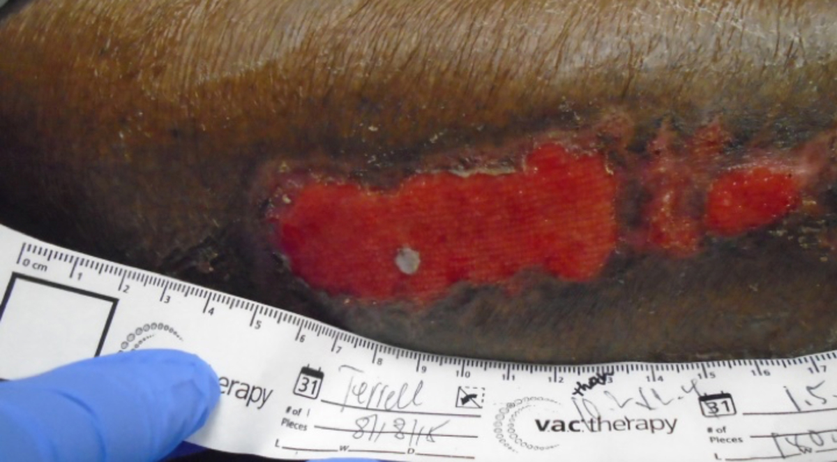
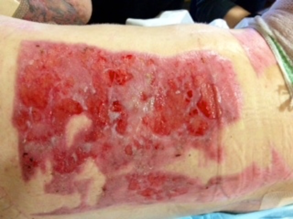
A 68-year-old African-American female underwent a below-the-knee amputation, due to severe peripheral arterial disease, and needed a skin graft to close the amputation. Comorbidities included hemodialysis-dependent end-stage renal disease and type 2 diabetes mellitus. Her visiting nurse removed her film dressings early in the first postoperative week and reapplied petrolatum-impregnated gauze and tape (Figure 1). The patient developed a hematoma over part of the site, and the wound never healed despite aggressive local care, which included absorptive antimicrobial dressings, treatment of hypergranulation tissue with silver nitrate, and collagen products (Figures 2 and 3). After 3 months of wound care and minimal improvement, a decision was made to excise and primarily close the donor site (Figure 4).
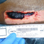 Figure 1. Donor site 1 week after graft harvest
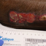 Figure 2. Donor site 1 month after graft harvest
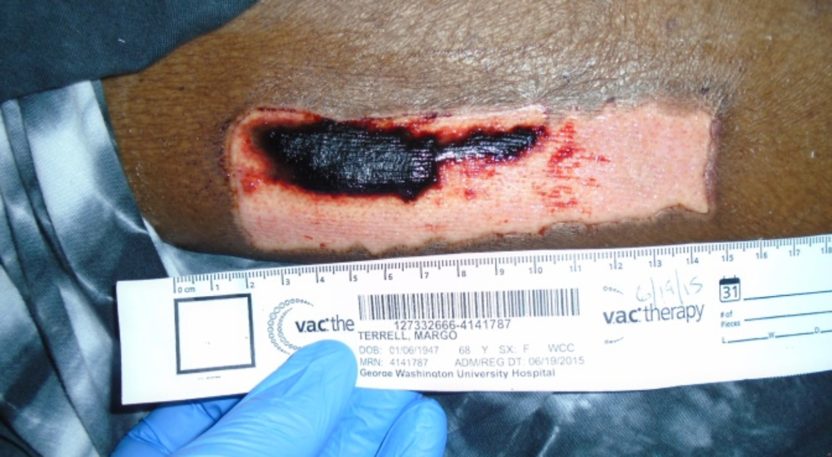
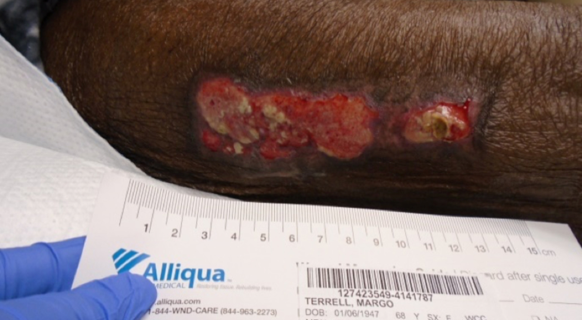
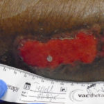 Figure 3. Donor site 2 months after graft harvest
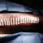 Figure 4. Delayed primary closure 3 months after graft harvest
CASE 2
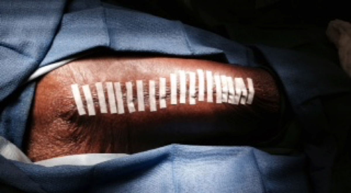
A 26-year-old Caucasian male with high bilateral above-the-knee amputations had donor sites on the flanks that became superinfected (Figure 5). Due to the amputations, the anterolateral thighs were not options for donor sites. The donor sites were colonized with multiple organisms over the course of his care as a trauma patient. Copious amounts of serous drainage exuded from the donor sites daily, and the sites were so painful that the patient needed dressing changes in the operating room three times a week. He was treated with systemic antimicrobials and then a contact layer (Figure 6). Hydroconductive dressings were used for pain control and exudate management (Figure 7). The patient was able to tolerate wound care at the bedside after a few weeks (Figure 8). Several months after grafting, donor site closure was achieved.
MANAGING DELAYED DONOR SITE HEALING
The following management strategies may be helpful if the donor site wound does not follow the expected course of re-epithelialization.
1.Reduce bioburden: Use an antiseptic compress, such as dilute acetic acid, 0.125% Dakins solution, or hypochlorous acid, for at least 20 minutes with each dressing change.
2.Use an advanced dressing: If the initial dressing did not produce a healed wound, change to a more advanced dressing, such as a polyurethane foam or a CMC hydrofiber; these dressings can manage increased amounts of exudate and can also be changed atraumatically on the wound bed.
3.Apply a contact layer: A contact layer is also appropriate for pain control if more absorptive dressings are needed.
4.Optimize nutrition: If there is delayed healing of a donor site wound, it is critical to optimize nutrition and provide supplementation when indicated.
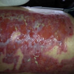 Figure 5. Chronic, nonhealing donor site on back/flank
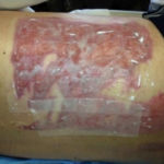 Figure 6. Contact layer.
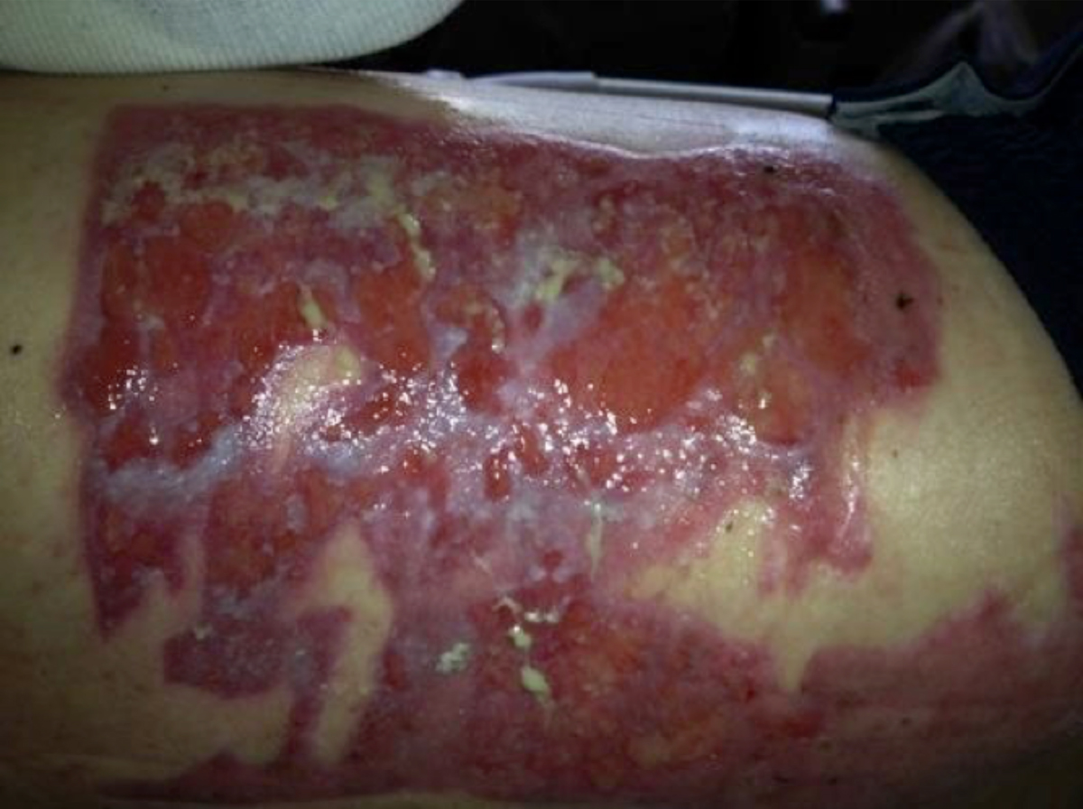
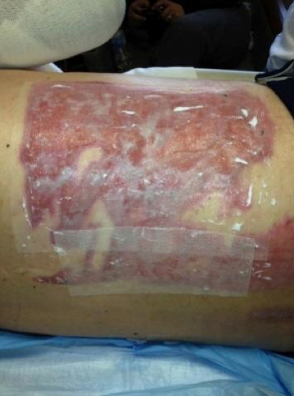
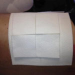 Figure 7. Absorptive secondary dressing
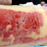 Figure 8. Gradual re-epithelialization. Bedside dressing changes were manageable after a few weeks.
CONCLUSION
In summary, donor site wounds can be hugely problematic for surgeons or wound care clinicians if the wounds do not heal within a few weeks of harvesting. A multimodal, aggressive approach is indicated to heal these wounds. If expected outcomes are not achieved, clinicians should look beyond the traditional dressings and consider using a composite dressing or an antimicrobial dressing to improve healing times. Education of the patient and nursing staff on troubleshooting dressings if problems develop can improve the likelihood of healing.
NOTE: As with any case study, the results and outcomes should not be interpreted as a guarantee or warranty of similar results. Individual results may vary depending on the patient’s circumstances and condition.
References
1.Voineskos SH, Ayeni OA, McKnight L, et al. Systematic review of skin graft donor-site dressings. Plast Reconstr Surg. 2009;124(1):298-306.
2.Dornseifer U, Lonic D, Gerstung TI, et al. The ideal split-thickness skin graft donor-site dressing: a clinical comparative trial of a modified polyurethane dressing and aquacel. Plast Reconstr Surg. 2011;128(4):918-24.
3.Brolmann FE, Eskes AM, Goslings JC, et al. REMBRANDT study group. Randomized clinical trial of donor-site wound dressings after split-skin grafting. Br J Surg. 2013;100(5):619-27.
4.Karlsson M, Lindgren M, Jarnhed-Andersson I, et al. Dressing the spilt-thickness skin graft donor site: a randomized controlled clinical trial. Adv Skin Wound Care. 2014;27(1):20-5.
5.Lauchli S, Hafner J, Ostheeren S, et al. Management of split-thickness skin graft donor sites: a randomized controlled trial of calcium alginate vs. film dressing. Dermatology. 2013;227(4):361-6.
6.Caliot J, Bodin F, Chiriac S, et al. Split thickness skin graft donor site: which dressing to use? Ann Chir Plast Esthet. 2015;60(2):140-7.
7.Geary PM, Tiernan E. Management of split thickness skin graft donor sites- results of a national survey. Clin Plast Surg. 2012;39(1):77-84.
8.Demirtas Y, Yagmur C, Soylemez F, et al. Management of split-thickness skin graft donor sites: a prospective clinical trial for comparison of five different dressing materials. Burns. 2010;36(7):999-1005.
