
Michael Robinowitz, M.D., earned his medical degree from Chicago Medical School in Chicago, Ill. He completed a residency and fellowship in obstetrics and gynecology at Emory University School of Medicine.Dr. Robinowitz joined Piedmont in 2009 as the medical director of the Wound Care Center in Fayetteville. Prior to joining Piedmont, he was in private practice at Nova OB/GYN in Atlanta and served as a Clinical Assistant Professor at Emory School of Medicine and Attending Faculty Physician at Grady Health System’s GYN/OB Clinic. He previously was in practice at Meridian Medical Group-Prucare of Atlanta where he served for some time as the President and Chairman of the Board.A fellow of the American Professional Wound Care Association and a diplomat of the American Board of Obstetrics and Gynecology, Dr. Robinowitz is board certified in obstetrics and gynecology and certified in wound care.
Robinowitz_Current Dialogues in Wound Management_2015_Volume 1_Issue 3
PATIENT
A 43-year-old male complained of pain, erythema, and swelling in his right groin that had persisted for 1 week. Initial care included antibiotics, aseptic skin cleanser, warm compresses, and incision and drainage of the abscess. Seven days later, the patient presented to the emergency room with a fever and increased right groin erythema, edema, and tenderness. Patient comorbidities included a history of smoking.
DIAGNOSIS
The patient was admitted with a soft tissue infection, abscess, and sepsis. Upon presentation to the emergency room, physical examination found right groin swelling, erythema, and tenderness. A small punctate opening in the right lateral suprapubic area was observed, with marked erythema down to the inguinal ligaments. Necrotic skin and marked fluctuance were present. A computed tomography (CT) scan of the pelvis indicated an elongated skin defect or wound in the upper anterior right thigh with extensive subcutaneous inflammatory changes, with nonloculated fluid densities, edema, and extensive gas bubbles extending from 20 cm below the hip joint to the right inguinal region and the base of the scrotum. Necrotizing fasciitis could not be ruled out.
INITIAL TREATMENT
The patient was taken to the operating room for exploration and surgical debridement of necrotic skin, subcutaneous tissue, and muscle tissue in the right thigh (Figure 1). The postoperative diagnosis suspected necrotizing fasciitis of the right thigh up to, but not involving, the scrotum. The wound was irrigated via pulsed lavage and packed with clorpactin-soaked gauze. Hyperbaric oxygen (HBO) therapy was started. An additional surgical debridement and irrigation with pulsed lavage was performed due to progression of necrotizing infection into the posterior and distal thigh (Figure 2). Clindamycin was added to the intravenous (IV) antibiotics.
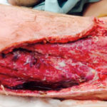 Figure 1. Right thigh wound following surgical debridement and wet-to-moist clorpactin-soaked gauze (postoperative day 1).
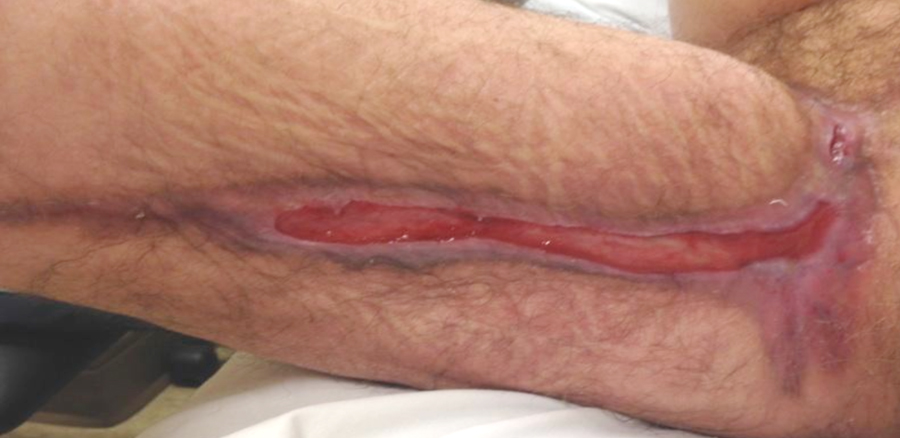
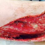 Figure 2. Right thigh wound following 2 hyperbaric oxygen treatments and minimal sharp debridement of loose nonviable tissue during wet-to-moist clorpactin gauze dressing change; wound measured 29 x 24 x 2 cm with a 2.5-cm tract and 4.2-cm undermining (postoperative day 2).
HBO treatments were continued, and the infectious disease service was consulted. Systemic antibiotics were continued, and a peripherally inserted central catheter (PICC) line was ordered. Dressings were changed daily with wet-to-moist gauze soaked with clorpactin solution. The PICC line was placed and antibiotics were continued
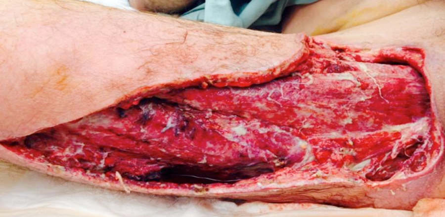
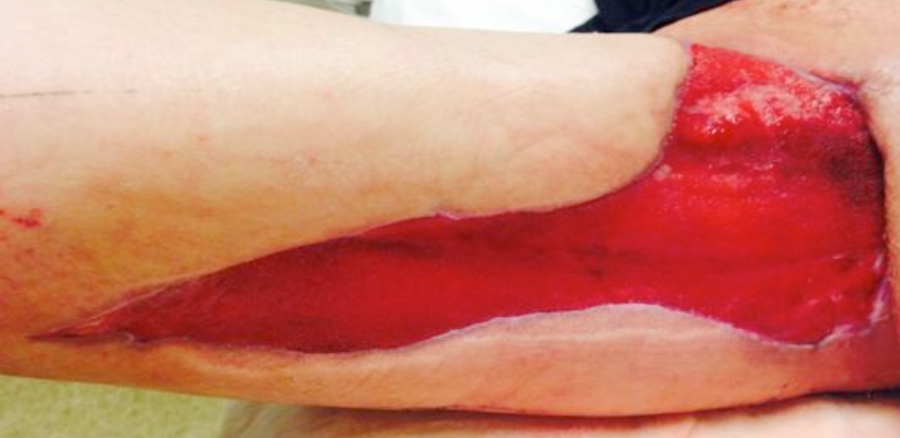
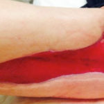 Figure 3. Right thigh wound following 21 days of negative pressure wound therapy; 100% granulation tissue and wound size reduction to 25.5 x 20 x 0.3 cm was observed (postoperative day 26).
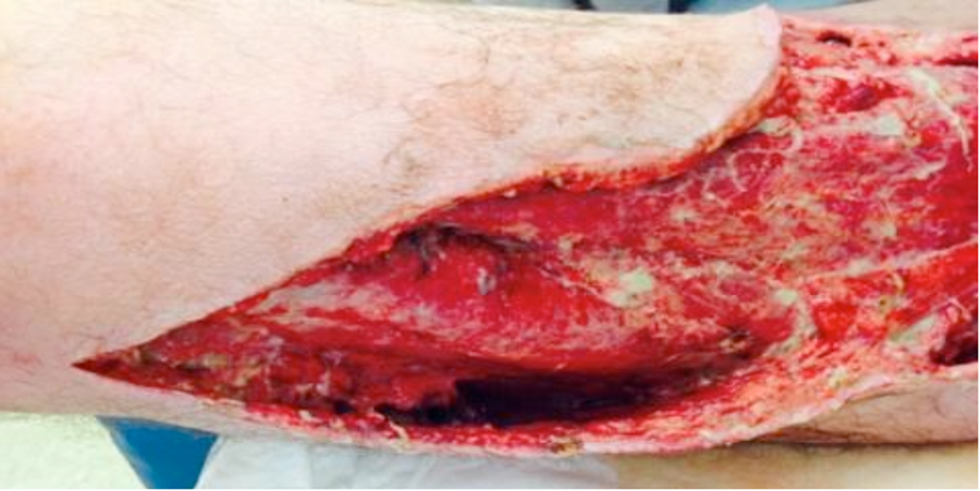
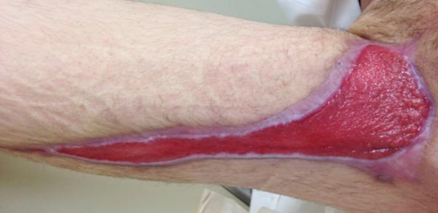
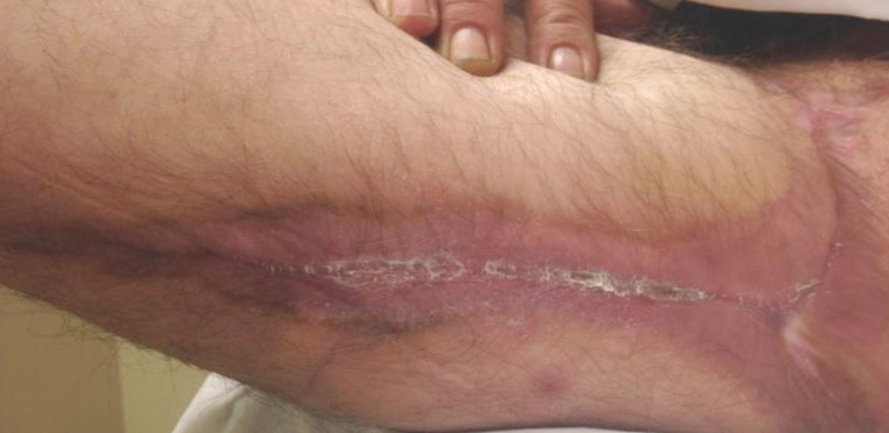
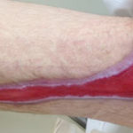 Figure 4. Right thigh following use of silver alginate dressing (postoperative day 42). New epithelium formation is visible around all wound margins.
APPLICATION OF V.A.C.® THERAPY
Negative pressure wound therapy (NPWT) with V.A.C.® Therapy (KCI, an Acelity company, San Antonio, TX) was initiated 7 days following presentation to emergency. At the time of NPWT initiation, the wound measured 29 x 24 x 2 cm with a 2.5-cm tract and 4.2-cm undermining. The wound contained <10% yellow slough in the wound bed. Both black foam (V.A.C.® GranuFoam™ Dressing, KCI) and white foam (V.A.C.® WhiteFoam Dressing, KCI) were used; NPWT was set for continuous therapy at -125 mm Hg. The patient noted only procedural pain during dressing changes. Wound care staff changed the dressings three times a week.
DISCHARGE AND FOLLOW-UP
Nine days following presentation, the patient was discharged with a PICC line, IV antibiotic therapy, and NPWT with home healthcare for dressing changes three times a week. The patient completed 21 days of NPWT. Wound measurements significantly decreased, and increased granulation tissue formation was observed. At the time of NPWT discontinuation, the wound measured 25.5 x 20 x 0.3 cm with 100% bright red granulation tissue (Figure 3) and moderate serosanguineous drainage. The plan of care was changed to a silver alginate dressing with a gauze dressing bolster and self-adhesive wrap to be changed three times per week. At each follow-up visit, the wound size decreased, and there were no further signs of infection or wound colonization (Figure 4). The patient regained full range of motion, mobility, and gait without skilled therapy intervention. The patient continued smoking throughout treatment. At the last clinic visit, the wound measured 11.7 x 1.4 x 0.1 cm (Figure 5). The patient was discharged 3 months following the last clinic visit (Figure 6).
USER EXPERIENCE
HBO therapy in conjunction with NPWT (using both black and white foam) contributed to an excellent outcome for this patient, better than would have been expected without use of these therapies, which promoted granulation tissue formation and contraction of a very large wound with extensive undermining and tracking. NPWT was initiated following several rounds of HBO treatment and wound debridement. Pain was only noted during NPWT dressing changes, but this was adequately managed with preprocedural pain medication.
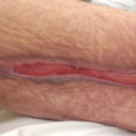 Figure 5. Wound healing by contraction with 100% granulation tissue; right thigh wound size reduced to 11.7 x 1.4 x 0.1 cm (postoperative day 105).
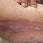 Figure 6. Right thigh wound completely healed 8 months following presentation to the emergency room.
CLINICAL OUTCOME
This case illustrates the successful use of NPWT in conjunction with appropriate antibiotic therapy, wound debridement, and HBO treatment. Following 21 days of NPWT, the large right thigh wound with undermining and tracking exhibited 100% granulation tissue. Upon discontinuation of NPWT, moderate serosanguineous draining was noted, and a silver alginate dressing was applied. No complications were observed, and the wound was completely healed 8 months following presentation to the emergency room without any need for skin grafting or other surgical closure.
NOTE: As with any case study, results and outcomes should not be interpreted as a guarantee or warranty of similar results. Individual results may vary depending on the patient’s circumstances and condition.
References
