
Jeffrey Litt, DO, is an Assistant Professor of Surgery and Medical Director of the Burn and Wound Program at University Hospital’s Frank L. Mitchell Jr., MD, Trauma Center. He joined MU’s Hugh E. Stephenson Jr., MD, Department of Surgery in July 2014. Born in Iowa but raised in Philadelphia, Dr. Litt completed undergraduate and medical school in Pennsylvania. He and his wife have two children.
Litt_Current Dialogues in Wound Management_2016_Volume 2_Issue 3
Burns, or acute traumatic wounds to the perineum or buttocks, are an infrequently suffered injury in the general population, but when they do occur, they can have serious and life-long consequences and may commonly contribute to injury morbidity. In the burn population, they most frequently occur in patients with larger surface area burns, but occasionally may occur in isolation in scald burns or in cases of abuse. There are limited data to evaluate the exact incidence of either burns or traumatic wounds to this region of the body; however, recently published burn literature at a busy North American burn center retrospectively put the incidence of buttock and perineal burns at approximately 25/year over a 3-year period (Merchant, 2014).1 In another review, the incidence of genital burns is estimated to be 2.8 – 13% (Michielsen, 2010).2Traumatic abrasions are likely even more commonly seen than burns given their association with motor vehicle accidents, particularly motorcycle-involved injuries. However, consistent data reporting, even in hospitalized patients, is lacking on these injuries.
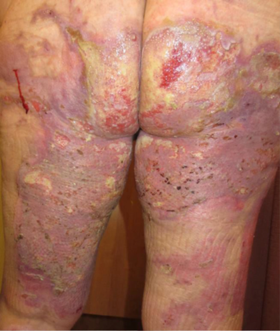
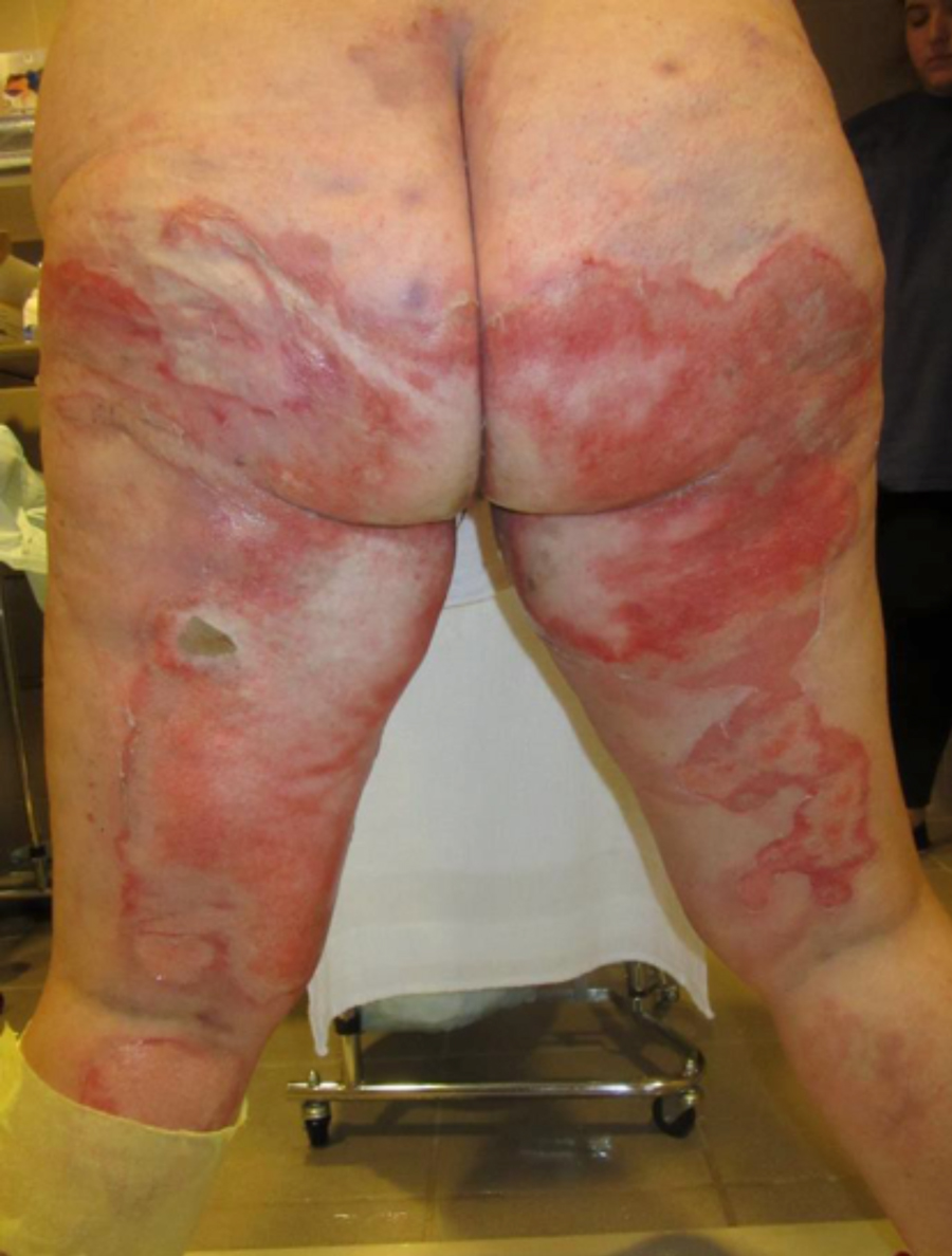 Figure 1. Initial presentation of buttock and posterior thigh burn wounds
Regardless of the type, injuries to this region are complicated and difficult to treat, primarily due to the difficulty maintaining dressings over the various curvatures and three-dimensional shape of the perineum/buttocks. In addition there is frequent contamination due to urination/defecation, as well as repetitive shear issues sustained during activity. Because of these challenges several controversies exist in the evaluation and management of buttock/perineal injuries. Initial dressing choice, need for fecal diversion, patient activity level, or timing of excision and choice of grafting comprise many of these controversies. Few studies have been performed evaluating these questions in the care of these patients, and much variation exists in specific burn/trauma unit protocols in dealing with these difficult wounds; these studies and their recommendations will be reviewed and evaluated. Finally, a review of a recently treated patient at our combined burn and trauma unit with significant buttock burns will ensue to review the concepts illustrated in this literature review.
In 1979, the US Military/US Army Institute of Surgical Research (USISR) reviewed its experience of 197 patients with perineal burns over a five-year period, noting a 71% mortality in patients with perineal burns – double the mortality compared with patients without perineal burns. Due to the high mortality and high incidence of complications, recommendations by the USISR working group included routine usage of systemic antibiotics, as well as surgical urinary diversion in cases of penile burns (Peck, 1990).3 In 1990, the burn team at the University of Washington compared its cases of perineal burns with those of the USISR and concluded that civilian mortality was significantly lower (28% vs 71%) and required less urinary catheterization and fewer surgical procedures (Peck, 1990).3Neither they nor the USISR discussed the need for fecal diversion, nor did they discuss specifics of wound care or patient mobilization.
Over 50 years ago, J Espinar reported on his experience in treating these wounds and suggested routine colostomy for fecal diversion to allow for appropriate wound healing (Merchant, 2014).1 In 2002, H Nakazawa et al reported on their experience treating five elderly patients with perineal burns using temporary diverting colostomy for assistance with wound management and cleanliness. They reported a mean patient age of 72 years old, a mean burn size TBSA of 30%, 2 mortalities (in the larger size burns, by their report unrelated to the perineal burns) and no ostomy-related complications. In fact, two of the patients had their colon continuity restored after their eventual recovery.
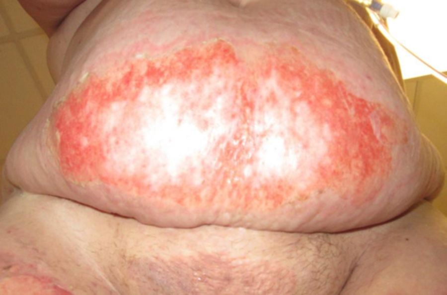
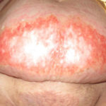 Figure 2. Initial presentation of pannus and suprapubic burns
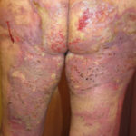 Figure 3. Split-thickness skin grafts hospital day 21, post-operative day 12
More recently, an evaluation and literature review by a group of French burn experts on perineal burn wound management (from eight different burn centers, average annual census of 219 patients, 13 average perineal burns/year) discussed usage of fecal management systems (FMS) rather than colostomy for fecal diversion as a first-line strategy. Nearly three-quarters (73%) of the burn centers stated they used a FMS for fecal diversion in sedated patients and over half (54%) in non-sedated patients without significant complication. In fact, they state that “the majority of patients who were alert and responsive had no pain or discomfort with the FMS in place” (Bordes, 2013).5 There is still a scarcity of data to support or refute the usage of these devices as a matter of routine; however, the evidence is mounting. The burn surgery group in Toronto reviewed its experience with buttock and perineal burns over a 3-year period: 74 patients were admitted over the period, of whom 13 were excluded, 12 died in the first 24 hours, and 1 was transferred before completion of treatment. Of the 61 patients they reviewed, only 5 received fecal diversion, and the remaining 55 received either a FMS (Flexi-Seal®, ConvaTec, Skillman, NJ) or routine wound care without specific fecal diversion. They subsequently reported operative procedures, procedures requiring revision, mortality, and infection rates. They found no statistical differences in any of the parameters measured between groups receiving FMS and ostomy creation (Merchant, 2014).
Acute wound care regimens for burns of the perineal region tend to be conservative. Daily cleansing and secured antibiotic dressings are the mainstay. Typically, even deeper appearing burns of perineal structures such as the penis or the labia respond well and heal within a 3-week period of time. In wounds not involving the anus, negative-pressure therapy can be very useful, not only as a barrier to fecal contamination/infection, but also as a bolster, allowing for earlier mobility with less shear concern. If negative pressure dressings are impractical or cannot be used, other bolster techniques, such as elastic outer dressings with tubular net bandages (Medichoice®, Owens & Minor, Mechanicsville, VA) or fashioning Montgomery Straps (Medline, Mundelein, Illinois) can be useful.
Traumatic wounds and abrasions of the perineal region are typically associated with motor vehicle accidents. In our experience, motorcycle accidents are the most commonly encountered cause of traumatic abrasions in this region. We have cared for a number of patients with buttock abrasions (approximately 20 over the last 14 months), and we have treated them all non-operatively. All of them healed completely, a majority in less than 3 weeks despite having the appearance of full-thickness wounds. They were all treated similarly, with judicious cleansing of the wounds followed by silver-based foam dressings for exudate management and infection prevention. Limited research is available on the incidence or routine care of these “road rash” type abrasions, but many consider them similar to, but more likely to heal than thermal injuries. Even full-thickness-appearing wounds tend to heal without need for skin graft if they are adequately cleaned and kept infection free. Regardless, in our combined burn and trauma service traumatic abrasions are commonly seen and, at least for the past 2 years, have never required operative excision or skin grafting.
Recently we cared for a patient with significant lower extremity and buttock burns and subsequently utilized many of the modalities previously mentioned. JM is a 50 year-old female admitted from the emergency department to the trauma/burn service with approximately 15%TBSA partial- and full- thickness burns to the bilateral buttocks, thighs, mons pubis, and pannus after her pants and shirt caught on fire (Figure 1,2). She was admitted and underwent operative burn wound debridement and xenograft placement approximately 1 day later. The following week she was brought back to the operating room for burn wound excision and autograft placement to the buttocks and thighs, along with primary excision and layered-closure of the pannus. We decided not to perform a fecal diversion procedure, given the presence of abdominal wall burns, as well the fact that she was managing to keep her dressings fairly clean and control her bowel movements. Skin graft dressings on the buttocks and thighs consisted of a non-adherent layer directly over the grafts, an elastic silver-based antibacterial layer that was stapled in place under tension, extra burn roll conformant and a compression layer of
elastic dressings (Medichoice). In addition, the patient was placed on a low-air loss mattress post-operatively to attempt to minimize graft shear. She was not kept prone. Dressings were taken off a week later with approximately 90% graft take, but there were areas of loss and shearing on the medial bilateral buttocks despite our shear-prevention attempts (Figure 3). Local wound care ensued, but by
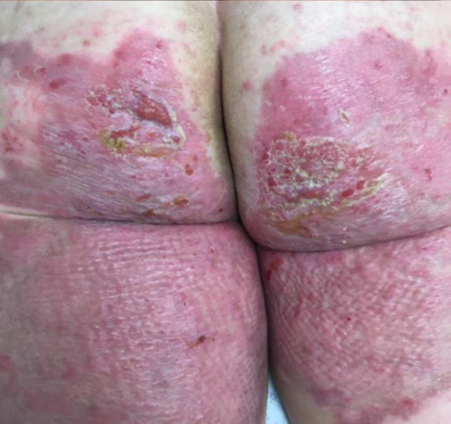 Figure 4. Final skin graft take post-operative day 56, 42
approximately 1 week later, it was clear the buttock wounds would need re-grafting. This was performed on hospital day 24, with a negative-pressure wound dressing utilized for graft protection. The grafts were fragile at dressing takedown 1 week later, requiring mafenide soaks and ultimately medical honey graft salvage. With local wound care, the patient’s wounds healed nearly completely approximately 1 month later, and the patient is currently undergoing scar management with custom compression garments (Figure 4).
Burns and wounds of this region are uncommon, but when they occur, they have a high likelihood of complications if care is inadequate. As a generalization, wounds should be cleansed free of debris and dressed with appropriate topical antibacterial agents, and the patients, especially if burns are present, should be referred to a burn center for further care and follow-up, per American Burn Association guidelines.
References
1.Merchant N, Boudana D, et al. Management of Adult Patients with Buttock and Perineal Burns: The Ross Tilley Burn Centre Experience. J Trauma Acute Care Surg. 2014;77(4):640-48.
2.Michielsen DP, Lafaire C. Management of Genital Burns: A Review. Intl J Uro. 2010;17:755-58.
3.Peck MD, Boileau MA, Grube BJ, et al. The Management of Burns to the Perineum and Genitals. J Burn Care Rehabil. 1990;11:54-56.
4.Nakazawa H, Ito H, Morioka K, et al. The Use of Diverting Colostomy to Manage Elderly Individuals with Extensive Perineal Burns. Burns. 2002;29:595-99.
5.Bordes J, Le Floch R, Bourdais L, et al. Perineal Burn Care: French Working Group Recommendations. Burns. 2014;40:655-63.
6.Huang T. (2012). Management of Burn Injuries of the Perineum. In Herndon, D (Ed), Total Burn Care: Fourth Edition (pp. 661-69). Elsevier.
7.Gottlieb L, Grevious M. (2006). Reconstruction of the Burned Perineum and Genitalia. In Sood, R (Ed), Achauer and Sood’s Burn Surgery Reconstruction and Rehabilitation (pp. 273-81).
