Dr. Bernstein maintains active surgical staff privileges as part of the Division of Podiatry / Department of Orthopedics within St. Luke’s University and Health System in Bethlehem, Pennsylvania, USA and is the Assistant Program Director of the podiatric surgical residency. He graduated from the Temple University School of Podiatric Medicine and completed a residency in foot surgery at Aria – Jefferson Health Bucks County Hospital, a Limb Salvage fellowship in Atlanta, Georgia, USA with Dr. Stanley Kalish, and a traveling fellowship in Limb Lengthening and Reconstructive Surgery with Ilizarov Technique at the Orthopedic and Traumatologic Department of Garcia de Orta Hospital in Almada, Portugal. Dr Bernstein is a paid consultant for 3M.
Bernstein_Current-Dialogues-in-Wound-Management_2023_Article-4
The patient was a 69-year-old male with a past medical history of non-insulin dependent diabetes mellitus, and ulcerative colitis who presented with a recalcitrant posterior leg ulceration overlying a necrotic Achilles tendon (Figure 1). The patient had a history of a ruptured Achilles tendon with primary repair in 1981; however, he stated that the wound had repeatedly opened and closed over the last 40 years. At the initial clinic visit, he noted increased and constant throbbing along with localized pain in this chronic wound, particularly at night.

Methods
After a lack of response over several months with conservative treatment consisting of zinc oxide paste wraps and local wound care by another physician, the patient was referred to our center for consideration of flap closure.
At the initial consultation, the wound was noted to have significant necrotic tissue throughout the undermined wound bed with extensive exposed devitalized Achilles tendon. The pre-flap workup included non-invasive vascular studies noting an ankle brachial index utilizing the peroneal artery of 1.41, metatarsal pressure of 212 mmHg and great toe pressure of 123 mmHg. A reverse sural artery flap was selected due to the location and size of the defect and presence of a patent peroneal artery. The reverse sural artery flap is a workhorse flap for the closure of soft tissue defects involving the distal third of the posterior lower leg and heel. As with all flaps, the reverse sural artery flap can have complications including venous congestion, arterial embarrassment, seroma or hematoma development, and infection.
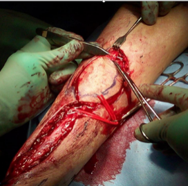
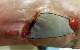
The sharp debridement and subsequent surgical closure were performed under general anesthesia. After thorough debridement, the wound measured 4.0 x 3.0 x 1.0 cm (Figure 2). Prior to flap incision, a sterile Doppler probe was used to map out the course of the small saphenous vein. The most distal perforator of the peroneal artery was identified in this manner approximately 5.0 cm from the distal tip of the lateral malleolus. The distance from the wound to the perforator was measured and included with the wound size so that the pedicle length and flap size could be templated. A reverse sural artery flap was dissected at the subfascial level from the posterior calf after ligation of the small saphenous vein, sural nerve and median sural artery and transposed onto the freshly debrided wound bed (Figure 3). A dressing with a bilayer bovine collagen/glycosaminoglycan/silicone graft was applied at the donor site with the plan of an eventual skin graft to that site. Post-surgical gauze wrapping was applied to both the donor site and the flap.
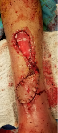
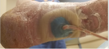
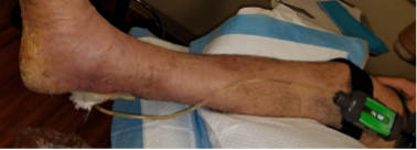
Upon post-operative follow-up, it was noted that the distal end of the flap progressively dehisced with the formation of unhealthy fibrotic tissue (Figure 4) and an undermined area was present that measured 6.0 x 7.0 cm (Shaded Area Figure 5). Therefore, the decision was made to return to the operative theater approximately four weeks after the initial surgery for hydrosurgical debridement of nonviable tissue and advancement and re-inset of the flap along with a split thickness skin graft (STSG) to cover the original flap donor site with a STSG from the ipsilateral calf. Due to the extensive undermining, supplemental negative pressure wound therapy was applied to bolster the flap and enhance healing of the open area of dehiscence. 3M™ Snap™ Therapy was selected due to the small nature of the wound and limited drainage as it would allow for better patient mobility, would be discrete/silent and could be worn under the patient’s clothes, would not require the patient to learn any complicated settings or controls, and would still allow application of 125 mmHg negative pressure utilizing an open cell reticulated foam construct (Figures 6-7). The cutoff in the author’s practice for use of Snap Therapy is a wound size less than 13.0 x 13.0 cm and drainage less than 180 mL/week.
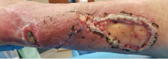
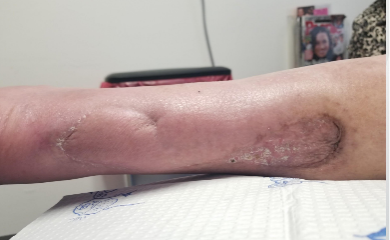
The patient was followed regularly post-operatively and had the Snap Therapy dressing changed twice weekly. Incremental reduction to the size of the open reticulated foam at each dressing change allowed slow collapse and healing of the undermining. Complete healing was noted at week 19 after the second surgery (Figure 8).
Conclusion
Surgeons must be cognizant of potential wound healing complications and plan for mitigation strategies. In this case, the retro Achilles area is well known for post-operative wound dehiscence due to the presence of limited blood supply in the watershed area. The presence of extensive non-pliable fibrotic soft tissue, undermining, and tenuous watershed blood supply all contributed to a recalcitrant non-healing wound of the Achilles region.
This case study highlights the successful use of Snap Therapy as an adjunctive treatment for salvaging a failing reverse sural artery flap. Snap Therapy gives the orthoplastic surgeon another tool to address post operative wound complications. Use of SnapTM Therapy can be of benefit in patients with a modestly active lifestyle who would not fare as well with use of the standard electrically powered negative pressure device.
References
NOTE: Specific indications, contraindications, warnings, precautions and safety information exist for these products and therapies. Please consult a clinician and product instructions for use prior to application. Rx Only.
As with any case study, the results and outcomes should not be interpreted as a guarantee or warranty of similar results. Individual results may vary depending on the patient’s circumstances and condition.
Patient data and images courtesy of Brent Bernstein, DPM.
©2023 3M. All rights reserved. 3M and the other marks shown are marks and/or registered marks. Unauthorized use prohibited. Used under license in Canada.
