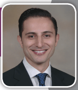
Dr. Tim Matatov completed his general surgery residency at Louisiana State University Health Shreveport and is board certified in general surgery. He is currently training in Plastic and Reconstructive Surgery at Tulane University. He has published in the Journal of Vascular Surgery and the American Journal of Surgery and was invited to be a guest editor for the International Wound Journal.
Matatov_Current Dialogues in Wound Management_2015_Volume 1_Issue 2
NO two wounds are the same; however, categorizing them and adhering to certain principles based on wound characteristics can certainly simplify and optimize their treatment. The broadest of these categories is the acute versus chronic wound. Acute wounds generally present with a brisk onset, and the four classic signs of inflammation – heat, pain, redness, and swelling. Chronic wounds, on the other hand, are insidious and rarely have these inflammatory changes. Differentiation is the key to treatment. Although acute wounds generally respond to systemic antibiotics and debridement, chronic wounds rarely respond to systemic antibiotics. Despite this, many practitioners start patients with chronic wounds on a prolonged course of antibiotics. This is not only futile, but encourages antibiotic resistance and unduly exposes patients to adverse effects from these medications. Why are systemic antibiotics ineffective in chronic wounds? It is because they are typically covered in biofilm.
Risk factors for biofilm formation in wounds include diabetic ulcers present for more than one month, a wound larger than 4 cm2, male gender, and previous antibiotic use.4 Biofilm is defined as any group of microorganisms that stick to each other on a surface and are embedded in a self-produced matrix of extracellular polymeric substance (EPS).5 Typically biofilm meets four criteria. First, attachment to a surface– whether it be a wound bed, suture, or an implant. This is the most powerful signal to initiate biofilm formation. Second, the bacteria secrete an EPS comprised of polysaccharides, host deoxyribonucleic acid (DNA), bacterial DNA, bacterial proteins, and host components. Biofilm can actually consolidate the components to tailor the EPS to protect itself from environmental threats. Third, a feature unique to biofilm is that it contains quorum-sensing molecules. These molecules function to direct gene expression of the different constituents of biofilm by creating a system of stimulus and response that reflects the population density within the biofilm. This explains why, within the often polymicrobial biofilm, different bacteria grow at different rates. Fourth, one of the peskiest features of biofilm is that it can essentially come back from the dead. Even if there is an event that destroys the majority of its constituents, the remaining pieces will combine and reconstitute. This period during which biofilm fragment reconstitution is taking place is when biofilm is at its most vulnerable, resulting in a therapeutic advantage for the practitioner.
Bacteria in wounds exist in two predominant forms: a free-floating, predatory, planktonic form typically seen in acute infections and a community of parasitic bacteria usually seen in chronic wounds. Diagnosing the presence of biofilm remains challenging and requires molecular methods (i.e., polymerase chain reaction [PCR]) to be done accurately.5-7 PCR findings should be confirmed with either imaging of bacterial biofilm or with culture techniques. Collecting wound cultures alone in chronic wounds is not helpful in improving wound outcomes and usually yields only the planktonic bacteria present in the wound bed. Proper identification of biofilm results in improved wound outcomes only if the appropriate therapy is initiated. A more rudimentary method of biofilm identification includes taking a biopsy of the wound and quantifying the number of colony-forming bacteria. If there are more than 105bacterial cells per gram of wound tissue, this is consistent with wound colonization and presumably biofilm formation. It is important to keep in mind that “105” is a general guideline, and as virulence of bacteria increases, the number for necessary bacterial load decreases. As mentioned earlier, systemic antibiotics are not effective in eradicating biofilm, and it must be approached with a combination of topical cidal agents, antibiofilm agents, and debridement. Sharp debridement is the most effective method to remove the majority of biofilm, and the remaining biofilm is more vulnerable and subsequently more susceptible to the host immune system and to topical cidal and antimicrobial agents.4,8,9 Topical cidal agents include povidone iodine and silver dressings. Anti-microbial agents include polyhexamethylene polyhexamethylene biguanide and octenidine with ethylhexyl glycerine biguanide. Although there is no definitive way to know if biofilm has been eradicated from a wound, clinical improvement is suggestive of successful therapy. Wounds will begin to heal and also produce less exudate.
SUMMARY
Biofilms are communities of bacteria, encased within a protective glycocalyx that functions to shield the bacteria from the host’s immune defenses. These parasitic colonies of bacteria contribute to nonhealing wounds and respond poorly to systemic antibiotics. Identification is best done with PCR and confirmatory wound culture. Once biofilms are identified, hallmarks of management include wound cleansing, sharp debridement, and topical cidal and antimicrobial agents. Wound healing and decreased exudate formation are indicative of successful therapy. Potential adjuncts in future management of biofilm include anti-quorum and biofilm-disrupting agents, which are currently in development.
References
1.Smith AW. Biofilms and antibiotic therapy: Is there a role for combating bacterial resistance by the use of novel drug delivery systems. Adv Drug Delivery Rev. 2005;10:1539-50.
2.Hoiby N, Bjarnsholt T, Givskov M, et al. Antibiotic resistance of bacterial biofilms. Int J Antimicrob Agents 2010;35(4):322–32.
3.Phillips P, Yang Q, Gibson D, Schultz G. Assessing the bioburden in poorly healing wounds. Wounds Intl. 2011;2(2):17–9.
4.Keast D, Swanson T, Carville K, et al. Ten top tips. Understanding and managing wound biofilm. Wounds Intl. 2014;5(2):20–4.
5.Wolcott R, Dowd S. The role of biofilms: are we hitting the right target? Plast Reconstr Surg. 2011;127:28S–35S.
6.Melendez JH, Frankel YM, An AT, et al. Real-time PCR assays compared to culture-based approaches for identification of aerobic bacteria in chronic wounds. Clin Microbiol Infect. 2010;16(12):1762-9.
7.Ehrlich GD. The need for PCR-based panel testing for syndromic infectious diseases. Mol Diagn. 1996;1(2):83–7.
8.Wolcott RD, Rumbargh KP, James G, et al. Biofilm maturity studies indicate sharp debridement opens a time-dependent therapeutic window. J Wound Care. 2010;19:320–8.
9.Phillips PL, Wolcott RD, Fletcher J, Schultz GS. Biofilms made easy. Wounds Intl. 2010;1(3):1-6.

