

Kris Bernaerts earned his Master of Science in Nursing in 1996. Since then, he has worked as a nurse at the Burn Center and the Intensive Care Unit of University Hospitals Leuven, where he also served as head nurse for several years. In 2004, he further specialized as a Clinical Nurse Specialist in wound care, a field to which he is deeply committed. He serves as vice-chair of the Wound Care Consultant Society of Belgium, a Flemish association for wound care. He regularly gives lectures and presentations both domestically and internationally on wound care and wound management. He is a guest lecturer at KU Leuven for students of Medicine, Pharmaceutical Sciences, and Nursing, as well as for nursing students at the university college in Leuven and Limburg. Internationally, he is actively involved in wound care projects in Africa. This included coordinating a wound care project at a regional hospital in the Democratic Republic of the Congo. In 2024, he wrote the handbook *Wound Management: The Complete Guide*, an indispensable resource for students, nurses, and physicians who wish to deepen or refresh their knowledge of wound care. The book will also be published by Elsevier UK in 2027. Kris Bernaerts is a paid consultant of Solventum.
Bernaerts_Current-Dialogues-in-Wound-Management_2026_Article-8
Introduction
The use of negative pressure wound therapy (NPWT) has evolved significantly over the past two decades and has become a widely used treatment method in modern wound care.1,2 This therapy is used for hard-to-heal wounds, such as diabetic foot ulcers and pressure ulcers, but is also applied in the management of surgical wounds, traumatic injuries, open abdominal wounds, sternal wounds, partial-thickness burns, and skin grafts.1–3
NPWT, when used on open wounds, is intended to create an environment that promotes wound healing by secondary or tertiary (delayed primary closure) intention by preparing the wound bed for closure, reducing edema, promoting granulation tissue formation and perfusion, and by removing exudate and infectious material. Collectively, these effects contribute to wound healing or efficient wound bed preparation for surgery, thereby helping to reduce treatment duration, hospital stays, and healthcare costs.1–3
Despite these benefits, the application of NPWT is often perceived as complex and time-consuming. The procedure involves several steps, including protecting the wound area, cutting the foam dressing to size, securing an airtight seal with an adhesive drape, and correctly positioning the negative pressure port. Additionally, dressings typically need to be changed two to three times a week to facilitate wound therapy and help prevent excessive tissue adherence or ingrowth into the foam.4
The Solventum™ V.A.C.® Peel and Place Dressing has recently been introduced into the wound care landscape as a simplified application of NPWT. This all-in-one integrated NPWT dressing makes it possible to apply therapy quickly and easily. It is suitable for a wide range of wounds, including chronic wounds, acute wounds, traumatic wounds, subacute and dehisced wounds, partial-thickness burns, venous ulcers, pressure ulcers, diabetic foot ulcers, flaps, and grafts. The dressing can be used on wounds with a maximum depth of 6 cm and undermining up to 2 cm, provided tunnelling is not present.5,6
The following case study highlights key considerations for the use of SolventumTM V.A.C.® Therapy with V.A.C.® Peel and Place Dressing.
Case study
A 22-year-old woman was admitted for surgical treatment of hidradenitis suppurativa (HS) involving the sternal region. Her medical history included corneal surgery more than 10 years earlier.
Deeply disseminated HS tunnels within the dermis were observed, with purulent discharge present during the excision procedure. After debridement, the wound measured 11.0 cm x 6.5 cm with a depth of 1.7 cm. Immediately following surgical excision of the HS-affected tissue, V.A.C.® Therapy was initiated using the Solventum™ V.A.C.® Granufoam™ Dressing and continuous -125 mmHg pressure. Following surgical removal of HS-affected tissue, the patient’s care was managed by a multidisciplinary wound care team.
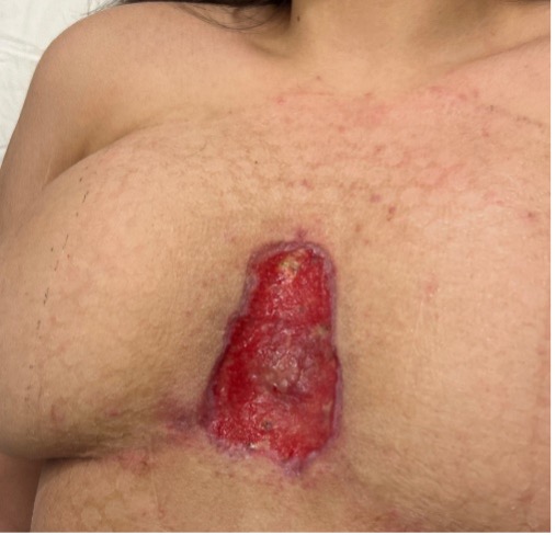
At the first dressing change 3 days later, (Figure 1), the patient reported severe pain with a Numerical Rating Scale (NRS) pain score of 10. A 2% topical lidocaine solution was applied to improve comfort during dressing removal.
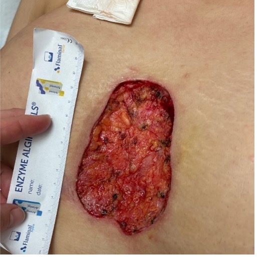
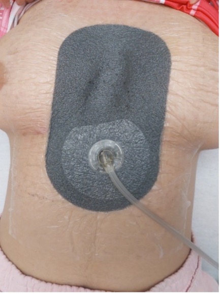
The wound care team decided to continue V.A.C.® Therapy but switched to the V.A.C.® Peel and Place Dressing to minimize pain during subsequent dressing changes. A medium-sized dressing was selected based on the wound dimensions. The integrated dressing and drape were applied over the wound and surrounding soft tissue (Figure 2). Continuous negative pressure of -125 mmHg was applied.
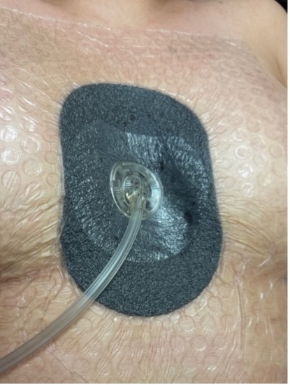
After 6 days of treatment, the first dressing change was performed. The patient reported less pain, with an NRS score of 4. The wound depth had decreased to approximately 1 cm (Figure 3).
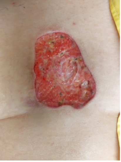
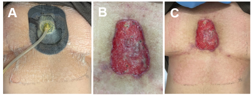
After a further 7 days of treatment, the second dressing change revealed dressing saturation due to pooling of wound exudate (Figure 4A). The negative pressure setting was not adjusted, but given the increased exudate, it might have been beneficial to increase the pressure to -150 mmHg. The wound showed a slight reduction in surface area to 9.0 cm x 6.5 cm and a depth of 0.6 cm. Wound edge advancement was observed, and healthy pink granulation tissue formation was noted, with a slight fibrinous or proteinaceous appearance (Figure 4B). This differed from the beefy red granulation tissue commonly observed when traditional NPWT dressings are applied directly to the wound bed.
No pain was reported during the dressing change. Furthermore, after removing the dressing, adhesive residue from the incorporated drape was observed along the edges of the application area. This is likely due to the adhesive strength of the drape and the length of wear time. The adhesive residue was easily removed using saline and did not cause any patient discomfort. Nevertheless, the dressing remains easy to apply and reposition during the initial dressing application. A temporary imprint from the drape was also visible (Figure 4C). This resolved spontaneously following discontinuation of therapy and was not considered clinically significant.
Therapy was reapplied and after a further 3 days, the dressing was removed due to the presence of an unpleasant musty odor (Figure 5). Despite this observation, the wound had continued to decrease in size to approximately 5.0 cm. Wound cultures were obtained, and while awaiting the results, the wound was treated with isobetadine. No microbial growth was detected and V.A.C.® Therapy was restarted 2 days later. A large V.A.C.® Peel and Place Dressing was used to improve the placement of the Solventum™ SensaT.R.A.C.™ Pad (Figure 6). Extending the SensaT.R.A.C.™ Pad outside the wound helps reduce the risk of skin irritation or temporary tissue deformation.
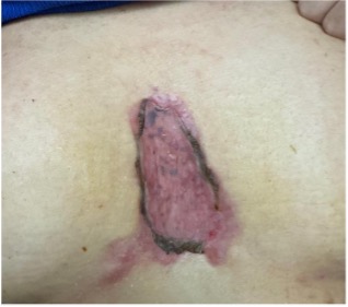
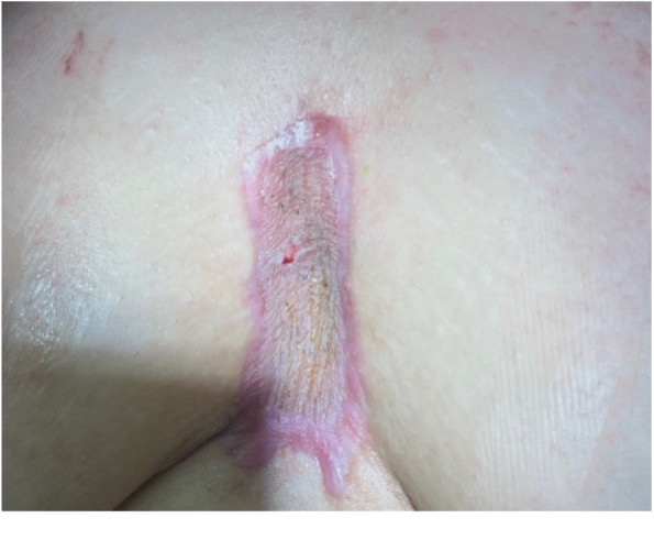
Following removal of the V.A.C.® Peel and Place Dressing in the operating room after an additional 6 days, the wound had nearly granulated completely, measuring 8.0 cm x 4.0 cm. After only four dressing changes over a four-week period, split-thickness autografts were applied. The skin grafts were covered with a monofilament non-adherent contract layer and a paraffin gauze dressing with dressing changes every 3 days. Follow-up at 3 days (Figure 7) and 3 weeks (Figure 8) after graft placement showed continued wound healing, with excellent graft adhesion and generally favorable healing.
Clinician experience
Application of the V.A.C.® Peel and Place Dressing was straightforward, requiring approximately one minute to apply and even less time to remove. Following transition to the V.A.C.® Peel and Place Dressing, the patient reported lower pain scores, from an initial NRS of 10 with the traditional V.A.C.® GranufoamTM Dressing. The incorporated Solventum™ Dermatac™ Drape allowed the V.A.C.® Peel and Place Dressing to be repositioned at first application; however, adhesive residue was observed after one week of wear, which was easily removed, without any discomfort to the patient. The clinical nurse specialist noted that the granulation tissue appeared more uniform. This differed from what the clinical nurse specialist had commonly observed with traditional reticulated open cell foam dressings. For this patient, the perforated, non-adherent layer of the V.A.C.® Peel and Place Dressing allowed for a longer wear time of up to 7 days, and the dressing could be removed with minimal to no pain.
References
-
Shehata M, Bahbah E, El-Ayman Y, et al. Effectiveness of Negative Pressure Wound Therapy in Patients With Challenging Wounds: A Systematic Review and Meta-analysis. Wounds. 2022;34(12):e126-e134. doi:10.25270/wnds/21061
-
Janssen AH, Wegdam JA, Reilingh TS de V, Eskes AM, Vermeulen H. Negative pressure wound therapy for patients with hard-to-heal wounds: a systematic review. J Wound Care. 2020;29(4):206-212. doi:10.12968/jowc.2020.29.4.206
-
Huang C, Leavitt T, Bayer LR, Orgill DP. Effect of negative pressure wound therapy on wound healing. Curr Probl Surg. 2014;51(7):301-331. doi:10.1067/j.cpsurg.2014.04.001
-
Morykwas MJ, Argenta LC, Shelton-Brown EI, McGuirt W. Vacuum-Assisted Closure: A New Method for Wound Control and Treatment. Ann Plast Surg. 1997;38(6):553-562. doi:10.1097/00000637-199706000-00001
-
Allen D, Mann S, Robinson T, Schmidt M, Kieswetter K. Preclinical Assessments of a Novel Peel and Place Extended-Wear Negative-Pressure Wound Therapy Dressing for up to 35 Days in a Porcine Model. Adv Wound Care. 2024;13(6):291-307. doi:10.1089/wound.2023.0096
-
Greenstein. Use of the new integrated 3M V.A.C. Peel and Place dressing on a diabetic foot ulcer. Current Dialogues in Wound Management. Dialogues in Wound Management. 2024:1-3. https://dialoguesinwoundmanagement.com/use-of-the-new-integrated-3m-v-a-c-peel-and-place-dressing-on-a-diabetic-foot-ulcer/
Patient data and images courtesy of Kris Bernaerts, MSc, RN.
NOTE: Specific indications, contraindications, warnings, precautions and safety information exist for these products and therapies. Please consult a clinician and product instructions for use prior to application. Rx only.
As with any case study, the results should not be interpreted as a guarantee or warranty of comparable results. Individual results may vary depending on the patient’s circumstances and condition.
©Solventum 2026. Solventum and S logo are trademarks of Solventum or its affiliates. 3M and the 3M logo are trademarks of 3M. All other trademarks are owned by their respective owners.